
The outlook in acute lymphoblastic leukemia (ALL) depends on the patient’s age, the biology of the leukemic cells, and above all on how completely the disease responds to treatment. These factors are combined into risk groups (risk stratification) that decide how intensive the therapy should be.
Childhood versus adult outcomes
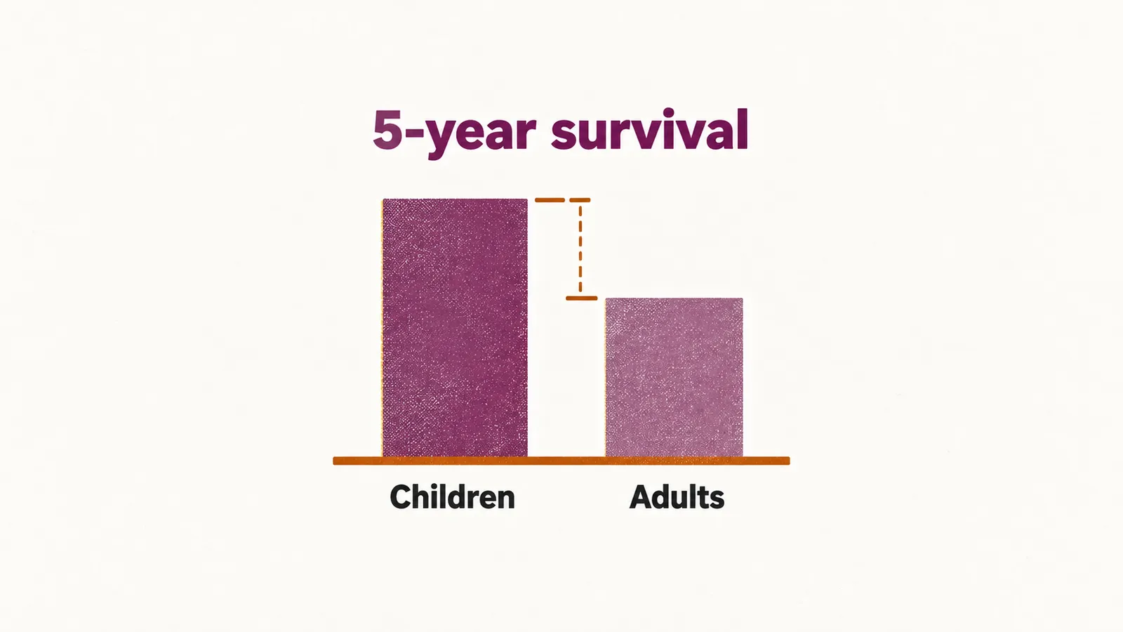
There is a large difference between age groups. About 90% of children with ALL survive at least five years, and most — roughly 80% — are cured, whereas 5-year survival in adults is about 30–40%. Across all ages the figure is about 72%, reflecting the high proportion of children among all patients. The gap reflects both differences in disease biology and the greater difficulty adults have tolerating intensive treatment.
Patient features: age and white cell count
The classic prognostic features at diagnosis are the patient’s age and the white blood cell (WBC) count. Children between 1 and 9 years of age do best; infants under 1 year and older adults do worst. A high WBC at diagnosis is adverse, and risk schemes use thresholds of about 50,000/µL in children and 30,000/µL in adults to separate standard- from higher-risk disease. The time taken to reach complete remission (the clearing of blasts from the marrow with recovery of normal blood counts) also carries weight: the shorter it is, the better.
Disease features: cytogenetics and immunophenotype
Cytogenetics, the chromosomal and genetic changes in the leukemic cells, carries prognostic weight. Some groups are favorable and others adverse:
| Outlook | Genetic change |
|---|---|
| Favorable | Hyperdiploidy, an increase in chromosome number involving trisomies (three copies) of chromosomes 4, 10 and 17; the ETV6-RUNX1 fusion t(12;21) characteristic of childhood B-ALL |
| Adverse | BCR-ABL1 t(9;22); rearrangement of KMT2A (formerly MLL) including t(4;11); hypodiploidy (a reduced chromosome number); complex or Philadelphia-like karyotypes |
The immunophenotype, the lineage a leukemia belongs to, also matters: B-lineage disease generally has a better outlook than T-lineage disease. Central nervous system involvement at diagnosis is adverse.
Response to treatment and minimal residual disease
The strongest single predictor is how the disease responds to treatment, measured as minimal residual disease (MRD) — leukemic cells present in numbers too small to see under the microscope, detected by flow cytometry or by polymerase chain reaction of immunoglobulin and T-cell receptor gene rearrangements. MRD that remains after induction predicts relapse, while MRD negativity — below about 0.01% of cells by flow cytometry, or below 10⁻⁴ by PCR — predicts lasting remission. MRD results are now used to adjust treatment intensity, for example by moving a patient towards stem cell transplantation, and they refine or override the older risk categories based on age, WBC count and cytogenetics. Relapse remains the main cause of treatment failure.