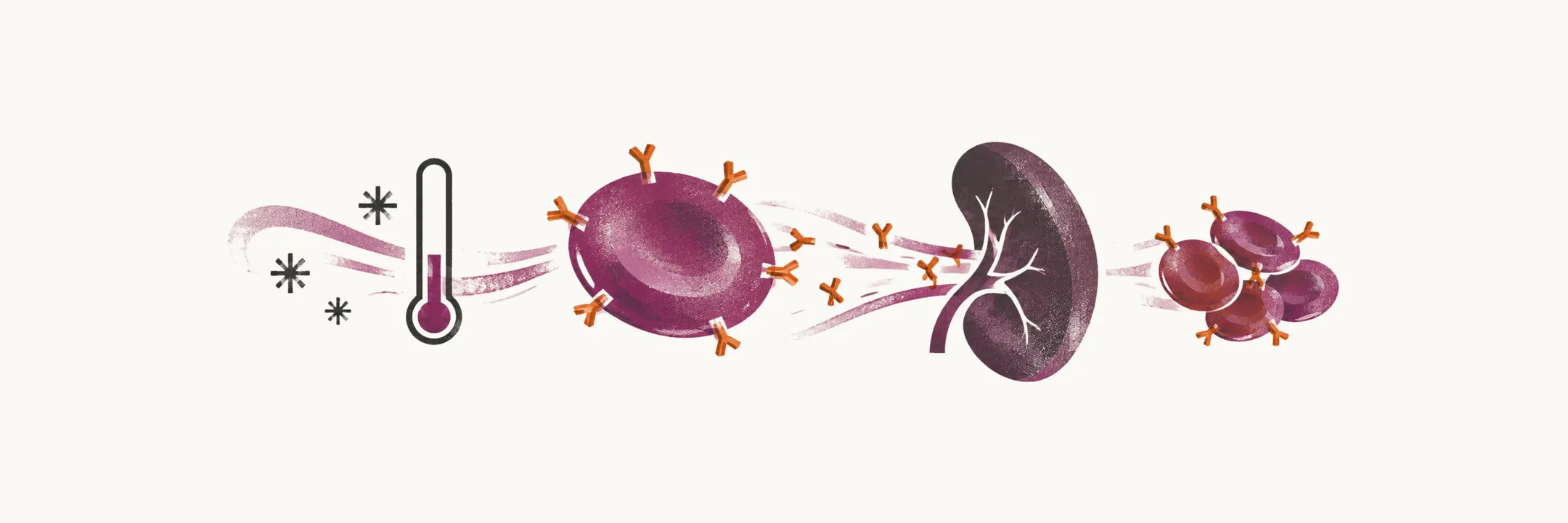
Immune hemolytic anemia has at least two distinct mechanisms, and the more frequent of them is a true autoantibody against the red blood cells.
Two immune mechanisms
- the innocent bystander mechanism, in which the immune system makes an antibody against another molecule, such as a drug, and red cells are caught in the resulting antigen–antibody complex;
- autoimmune hemolytic anemia (AIHA), in which a true autoantibody is directed against the red cells.
AIHA is the more frequent of the two, and it can be serious: even with appropriate management the mortality rate approaches 10%. It is uncommon overall, with an incidence of about 1 to 3 per 100,000 people per year. It is classified by the temperature at which the antibody reacts with the red cell:
- warm type autoimmune hemolytic anemia (WAIHA);
- cold type autoimmune hemolytic anemia (CAIHA).
Warm type autoimmune hemolytic anemia
The onset of WAIHA can be abrupt and acute. The picture is suspicious when a triad appears:
- an abrupt fall in hemoglobin, to as low as 4 g/dl;
- jaundice;
- splenomegaly.
Because the antibody reacts at body temperature, the coated red cells are cleared by macrophages in the spleen — the extravascular route — which is why the spleen enlarges. Reticulocytosis is usually present, because the marrow mounts a response, except when the autoantibody also targets the erythroid cells of the bone marrow. The general markers of hemolysis are found alongside it: increased LDH, decreased haptoglobin, and hemoglobinuria. In some patients, at the first presentation or later, autoimmune thrombocytopenia appears too, and the combination is called Evans syndrome.
The antibodies made against the red cells in AIHA are mostly Rhesus-specific autoantibodies. The Coombs test (the direct antiglobulin test) detects antibody and complement components attached to the red cell surface. A positive result means that antibody or complement is attached, which supports an immune cause, but a positive result alone does not prove that hemolysis is taking place.
The sensitivity of the test is limited: at least 300 to 400 molecules of antibody must be attached to each red cell for it to turn positive. A patient with a negative Coombs test can therefore still have AIHA, which can be severe if the affinity of the antibody is very high.
Treatment
Treatment almost invariably includes blood transfusion, and there is a paradox behind it: the transfused blood can be hemolyzed even when the ABO antigen is matched, because it is incompatible with the Rhesus-specific antibodies. Transfusion is still preferred in that situation, even with ongoing hemolysis, because the transfused blood acts as a reservoir while the patient’s own blood is continuously destroyed and the patient has to be kept alive.
If the hemolysis is not life-threatening, transfusion does not have to be used; prednisone or rituximab is used instead. Splenectomy is not a cure, but it can help by removing the major site of hemolysis.
Cold type autoimmune hemolytic anemia
Cold AIHA has a primary and a secondary form:
- primary — cold agglutinin disease (CAD);
- secondary — paroxysmal cold hemoglobinuria (PCH).
PCH is a rare cold AIHA that arises in children after a viral infection, is usually self-limited, and is characterised by the Donath–Landsteiner antibody, whose serology is distinctive: it attaches to red cells at 4 °C, and hemolysis follows when the temperature rises to 37 °C. In the body this produces large intravascular hemolysis.
CAD, the primary form, differs from every form of warm AIHA:
- it is chronic and more indolent, while WAIHA is more abrupt and acute;
- it reacts with red cells only at low temperature, which is why it is called cold;
- its antibodies are IgM, made by a clone of reactive B cells;
- its antibody is anti-I.
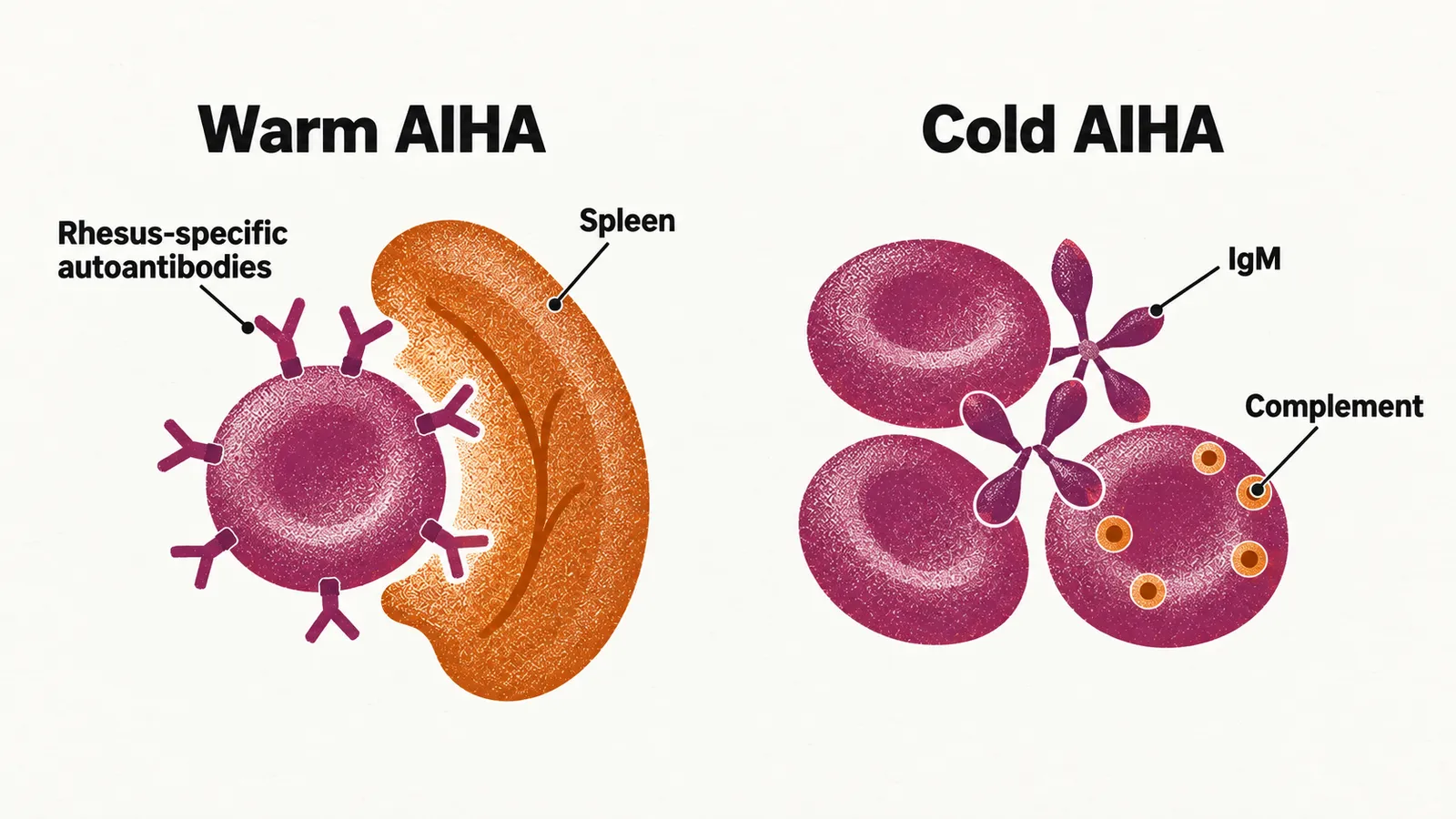
Because the antibody is IgM, the hemolysis proceeds in two ways:
- intravascular hemolysis — IgM causes direct membrane damage with the help of C9 of the complement system;
- extravascular hemolysis — C3b, an opsonin (a molecule that marks a cell for engulfment by phagocytes), marks the red cells for removal in the spleen.
Treatment is directed at the B-cell clone or at the complement system, and keeping the patient warm is part of management. Rituximab is the most accepted first-line therapy, and sutimlimab, an inhibitor of the complement protein C1s, was approved in 2022 for the hemolysis of CAD.
In CAD complement acts on red cells that are coated with antibody. Complement can also destroy red cells when the cell itself has lost the proteins that hold complement in check, with no antibody involved.