
Hemolysis is a shortened life span of the red blood cells, and it becomes hemolytic anemia only when destruction outruns what the bone marrow can replace.
Why a mature red cell cannot repair damage
Erythroid cells, the cells of the red cell line, differentiate from the stem cell line into fully mature red blood cells. During that process the cell accumulates hemoglobin in very high amounts while it degrades its organelles and loses most of its biosynthetic ability. The course looks self-destructive, but it produces a cell built for one job — carrying oxygen to the tissues for an average of 120 days — with almost no capacity to renew itself.
That is where the vulnerability lies. A mature red cell has a low metabolic reserve, so any alteration it sustains cannot be compensated, and its life span falls. The damage runs through one of two routes: structural damage to the cell membrane, or failure of the cation pump that maintains the cell’s ion and water content. Either route ends in the same place, a shortened red cell life span, which is what defines a hemolytic disorder.
From shortened life span to anemia
A hemolytic disorder does not by itself make the patient anemic. If the rate of destruction of red cells exceeds the rate at which the bone marrow produces new ones, the hemolytic disorder manifests as hemolytic anemia (HA). The common pathophysiologic pathway of every form of HA is therefore increased red cell turnover.
The reference method for measuring how long red cells survive is the red cell survival study, which follows labeled cells over time and shows the shortened life span directly.
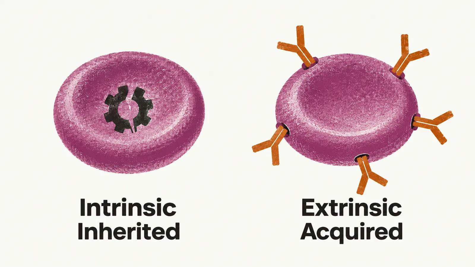
Where the defect sits decides which cells are destroyed, and this has a practical consequence. In an intrinsic (intracorpuscular) defect — a faulty membrane protein, enzyme, or hemoglobin molecule — the abnormality is built into the red cell, so the patient’s own cells carry it and the cause is usually inherited. In an extrinsic (extracorpuscular) defect — an antibody, a mechanical device, a toxin — the red cells themselves are normal and are destroyed by their surroundings, so the cause is usually acquired. Extrinsic hemolysis damages transfused red cells as readily as the patient’s own, so transfusion buys time rather than correcting the problem, whereas in intrinsic hemolysis only the patient’s defective cells are at risk.
Compensated hemolysis
When the kidney detects the fall in oxygen delivery it raises its production of erythropoietin (EPO), and the bone marrow expands red cell production in response. If that response keeps pace with the rate of destruction, the hemolysis is compensated and the patient is not anemic. A patient with hemolysis can therefore be non-anemic, which matters because the compensated state is not stable. Several conditions take away the marrow’s reserve and tip the patient into a decompensated, anemic state:
- pregnancy, which increases red cell demand;
- folate deficiency, which limits how fast the marrow can divide;
- renal failure, which interferes with EPO production;
- infection, which can interrupt red cell production.
Infection has a well-known example. Parvovirus B19 infects red cell precursors and halts their production for one to two weeks. A healthy person barely notices the pause, because red cells normally circulate for four months; a patient whose cells already survive only weeks loses the marrow’s output while destruction continues, and a rapid, severe aplastic crisis follows, usually requiring transfusion.