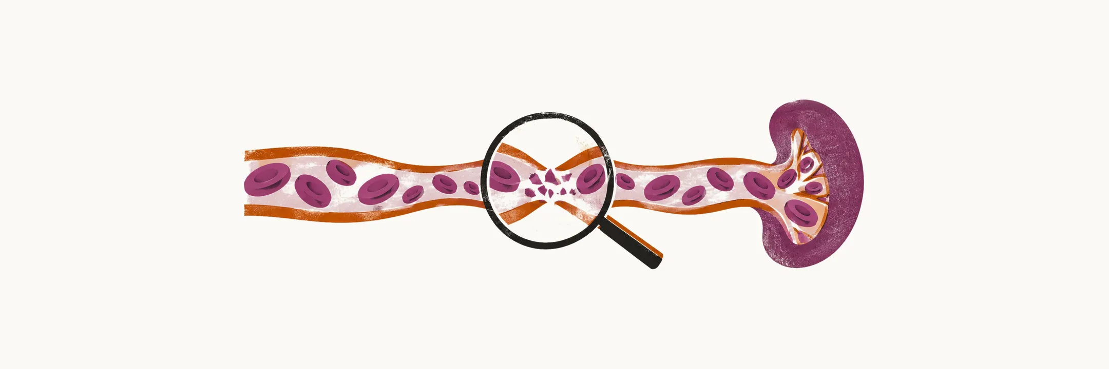
The signs and symptoms of hemolytic anemia come from the destruction of red blood cells and from the bone marrow’s response to it, and that combination is what separates it from the other anemias.
The clinical picture
The clinical presentation of the anemic patient, in hemolytic anemia (HA) as in every other anemia, is strongly influenced by whether the onset of signs and symptoms is abrupt or gradual. That part is shared. What distinguishes HA from the other anemias is a set of features produced by the red cell breakdown itself:
- jaundice, usually the leading clinical sign, from the unconjugated bilirubin released as red cells are destroyed;
- splenomegaly, present in many cases because the spleen is the preferential site of red cell destruction;
- hepatomegaly, which is less common than splenomegaly;
- skeletal changes, found in all severe congenital forms because the overactive bone marrow expands within the bone;
- discoloration of the urine, which appears when hemolysis is intravascular and free hemoglobin is passed in the urine.
The laboratory picture
The laboratory findings belong to two groups: markers of the hemolysis itself, and markers of the bone marrow’s response to it. The first group depends on where the red cells are destroyed.
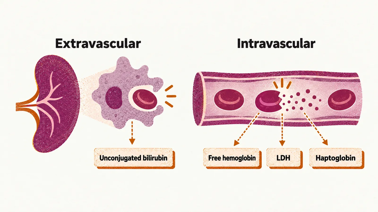
In extravascular hemolysis, red cells are engulfed and broken down by macrophages, mainly in the spleen, so free hemoglobin does not reach the plasma, but the breakdown products do:
- increased unconjugated bilirubin, the form of bilirubin produced from hemoglobin before the liver processes it;
- increased serum AST (aspartate aminotransferase);
- increased urobilinogen in both the stool and the urine.
In intravascular hemolysis, the red cells rupture inside the vessels and release free hemoglobin directly into the plasma:
- increased serum free hemoglobin, which mostly leads to hemoglobinuria;
- increased LDH (lactate dehydrogenase) in serum;
- decreased haptoglobin, the plasma protein that binds free hemoglobin and is used up in the process.
The bone marrow answers both patterns in the same way:
- reticulocytosis, an increase in reticulocytes, the young red cells released by the marrow;
- increased MCV (mean corpuscular volume, the average size of the red cells), which is due to the increased number of reticulocytes, since they are larger than mature red cells;
- erythroid hyperplasia in the marrow, an expansion of the red cell precursors.
Iron problems
Because hemolysis takes red cells out of the circulation, it disturbs the body’s iron balance, and the direction of the disturbance follows the site of destruction.
In chronic intravascular hemolysis, iron deficiency develops, because iron is lost from the body in the urine as hemoglobin. In chronic extravascular hemolysis, the opposite happens and iron overload develops. A patient with chronic extravascular hemolysis who needs blood transfusions runs a still higher risk of iron overload, but overload can develop without any transfusion at all: the secretion of erythroferrone rises, and erythroferrone inhibits hepcidin, the hormone that normally restrains iron absorption in the intestine, so iron absorption by the patient increases.
These features show that red cells are being destroyed and where the destruction takes place. They do not show why the cells are being destroyed, and that depends on whether the defect lies in the red cell itself or acts on it from outside.