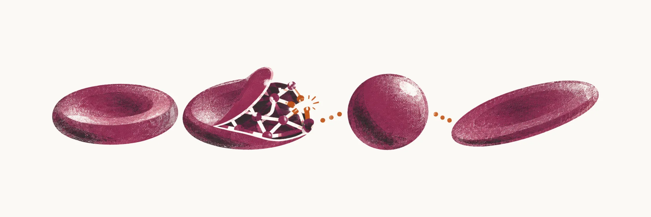
Defects of the red cell membrane and of the cytoskeleton that supports it are one of the inherited groups of hemolytic anemia, alongside the hemoglobinopathies and the defects of the cell’s channels and enzymes. The disorders that follow are easiest to understand from the structure that fails first.
How the membrane and cytoskeleton are built
The red cell membrane carries several classes of protein. Integral proteins extend internally and externally out of the membrane, while glycosylphosphatidylinositol (GPI)-anchored proteins extend only outside the cell. Functionally, the main membrane proteins are ion channels, energy-dependent ion transporters, complement receptors, and receptors for other ligands.
The most abundant membrane protein is band 3, an anion transporter. Its extracellular domains are heavily glycosylated and carry the antigenic determinants that correspond to the blood group.
Tangential to the membrane runs a cytoskeleton that contacts the membrane proteins. Its most abundant components are the spectrins, assembled into a spectrin tetramer by the head-to-head association of two α-spectrin–β-spectrin heterodimers. The cytoskeleton attaches to the membrane in two ways: through the ankyrin complex, which includes band 4.2, and through the junctional complex, which includes band 4.1.
This membrane–cytoskeleton complex is essential to the survival of the red cell, and a change in any part of it alters the cell’s deformability and ends in hemolysis. Almost all of these disorders are inherited diseases caused by mutations. Because the altered cell loses its shape before it lyses, an abnormal morphology is almost always visible on the peripheral blood film.
How the blood film sorts the disorders
Because morphology changes first, the common disorders of this group can be classified by the shape the film shows:
- hereditary spherocytosis — spherical cells;
- hereditary elliptocytosis — elliptical cells;
- stomatocytosis — mouth-shaped cells; this one also counts as a channelopathy, because the shape change comes from an ion-channel defect.
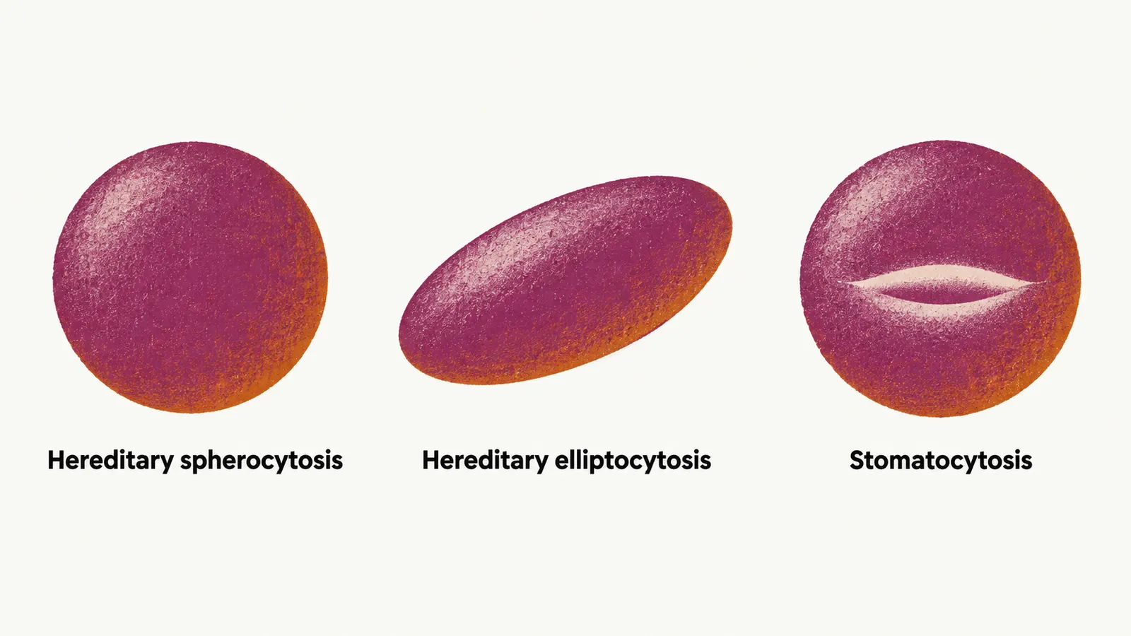
Most membrane–cytoskeleton disorders are monogenic, caused by a single gene, but there is no strict correlation between a single gene and a single disease: different mutations of the same gene can give rise to different disorders. Spherocytosis and elliptocytosis show this with proteins that sit in different positions in the membrane.
Hereditary spherocytosis
Hereditary spherocytosis (HS) is the most prevalent of these membrane disorders, found in 1 in 2000 to 1 in 5000 people of European ancestry. It is mostly caused by mutations in the band 3 gene and the ankyrin gene, and to a lesser extent the spectrin-β gene — all proteins that are oriented vertically in the membrane, linking the lipid bilayer to the cytoskeleton underneath. When those links weaken, the cell loses membrane surface relative to its volume and rounds up into a spherocyte, which is less deformable and is trapped and destroyed in the spleen.
That surface-to-volume mismatch is also why spherocytes are extremely susceptible to lysis in hypotonic media (solutions more dilute than the cell’s contents, into which water enters), a property called osmotic fragility. The classic osmotic fragility test is no longer the best single test: the eosin-5’-maleimide (EMA) binding test, measured by flow cytometry, is the recommended screening test together with cryohemolysis, and a normal osmotic fragility result does not exclude HS in 10 to 20% of cases.
HS is genetically very heterogeneous, since several different mutations in one of a group of genes can produce it, and it is not only autosomal dominant — autosomal recessive forms also exist. The clinical spectrum is wide: severe forms appear in infancy with severe anemia, while mild forms may appear in young adults or later.
The main clinical manifestations are:
- jaundice;
- splenomegaly;
- gallstone formation.
In the milder cases hemolysis is usually compensated, and anemia appears only under a challenge such as pregnancy or infection, in what is called the decompensated form. On the film the cells are spherical, so the central pallor of a normal red cell is not seen and the cells look hyperchromic (densely stained). The laboratory findings are a normocytic anemia when the hemolysis is not compensated, together with increased MCHC (mean corpuscular hemoglobin concentration), increased RDW (red cell distribution width, a measure of variation in red cell size), and a normal or slightly decreased MCV (mean corpuscular volume).
There is no treatment that repairs the membrane defect, but splenectomy reduces hemolysis by removing the main site of red cell destruction:
- in severe HS, splenectomy is recommended, ideally delayed until after the age of 6 years;
- in moderate HS, it is considered, towards puberty;
- in mild HS, it is best avoided.
Because the spleen also clears encapsulated bacteria, splenectomy carries a lifelong risk of overwhelming sepsis, particularly pneumococcal. Patients are vaccinated before surgery and are given prophylaxis according to local policy.
Hereditary elliptocytosis
Hereditary elliptocytosis (HE) is as heterogeneous as HS, with a prevalence of about 1 in 2000 to 1 in 4000. It is mostly due to a mutation in spectrin-α and, to a lesser extent, the spectrin-β gene and protein 4.1 — proteins that sit horizontally under the membrane, in the junctional complex. Most people with HE are asymptomatic, and only a minority develop uncompensated hemolysis. The number of elliptocytic cells on the film does not track the severity: some mild forms show almost 100% elliptocytes.
The most severe cases of HE, with severe hemolytic anemia, are those with biallelic mutations (mutations in both copies of the gene) in the membrane–cytoskeleton genes; they are called hereditary pyropoikilocytosis (HPP). Splenectomy, after vaccination, is the only specific treatment for the severe hemolytic forms of HE, and folate supplementation is advised while hemolysis continues.
Not every mutation in these genes causes disease. An in-frame deletion of 9 amino acids in the band 3 protein, for example, probably selected for by malaria infection, improves survival in some exposed populations; because it does not cause disease it is not called a mutation but a polymorphism, found mostly in southeast Asia and named southeast Asian ovalocytosis (SAO).
The third shape on the film, the stomatocyte, arises differently: the membrane is intact, but an ion-channel defect changes the cell’s volume.