
Channelopathies are defects of the ion channels of the red cell membrane, and they cause hemolysis by changing the volume of the red cell.
The volume problem
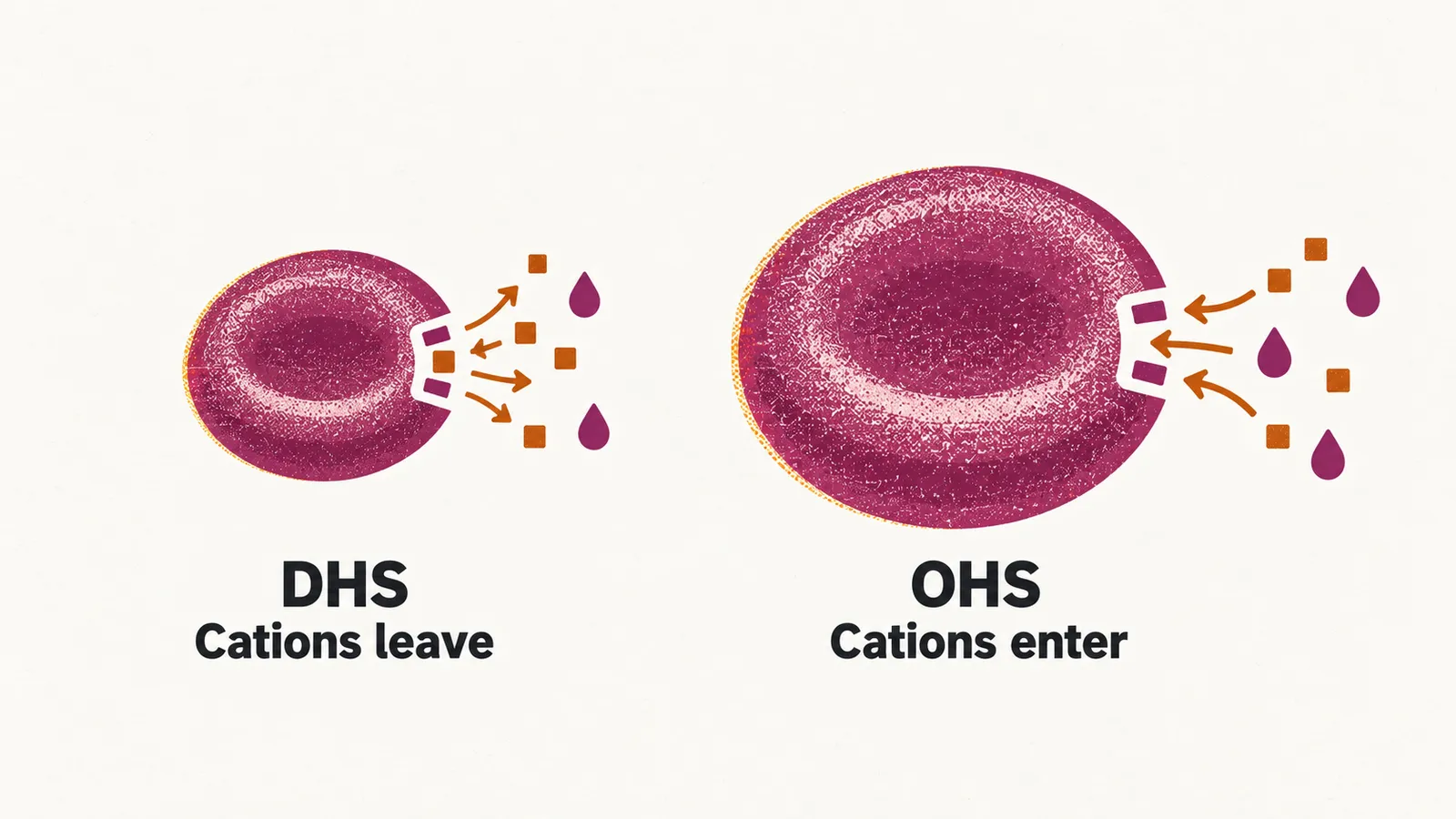
The main clinical feature of a channelopathy is a change in red cell volume, which follows from a change in the cell’s ion content: positively charged ions (cations) move across the membrane, and water moves with them. These are very rare causes of hemolytic anemia, and the two that matter are the stomatocytoses, named for the mouth-shaped stomatocyte seen on the blood film, which take a dehydrated and an over-hydrated form.
| Feature | Dehydrated hereditary stomatocytosis (DHS) | Over-hydrated hereditary stomatocytosis (OHS) |
|---|---|---|
| Ion movement | Cations leave the cell, and water follows | Cations enter the cell, and water follows |
| Cell water | Decreased | Increased |
| Why it is macrocytic | The cell raises its protein content to defend osmotic pressure, so it stays large despite the water loss | The water entering the cell expands it |
| Gene | PIEZO1, an ion channel activated by pressure (a mechanoreceptor); KCNN4, a calcium-dependent potassium channel | RHAG (the Rhesus gene), encoding an ammonia transporter; SLC2A1, encoding the GLUT1 channel |
DHS is the more frequent of the two, and it is a macrocytic hemolytic anemia, meaning that the red cells are larger than normal. The cation leak lowers the water content of the cell, and the compensatory rise in intracellular protein is what keeps the MCHC (mean corpuscular hemoglobin concentration) increased.
OHS is also a macrocytic hemolytic anemia, but its macrocytosis has the opposite cause: a higher cation content inside the cell pulls water in and expands it.
Why splenectomy is contraindicated
In hereditary spherocytosis (HS) the spleen is the main site of red cell destruction, and removing it reduces hemolysis. Hereditary stomatocytosis is the opposite case, and splenectomy is contraindicated. The abnormal cells need to be sequestered from the circulation; if the spleen is removed they stay in the blood, and thrombotic complications follow.
Cell volume depends on more than the channels: the cation pump that maintains the cell’s ion and water content also needs a steady supply of energy, which the red cell’s enzymes provide.