
A red cell depends on a small set of enzymes for its energy and for its protection against oxidation, and hemolytic anemia follows when one of them is deficient.
Two jobs, two time courses
Enzyme abnormalities cause hemolytic anemia when a deficient enzyme leaves the red cell unable to do one of its two essential jobs:
- producing ATP, the cell’s energy currency;
- maintaining its red-ox (oxidation–reduction) balance.
The prevalence of hemolytic anemia from an enzymopathy is very low, so it is considered only after other causes have been excluded. The two groups differ in how they present, and the reason lies in the job the enzyme performs.
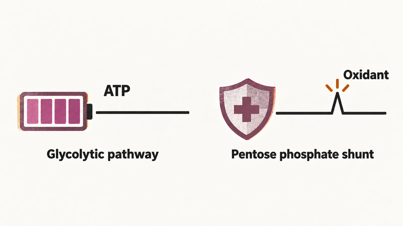
The pentose phosphate shunt protects the cell against oxidative damage, a threat that arrives in episodes; a defect there therefore tends to cause acute hemolysis, with nothing happening until an oxidant appears. The glycolytic pathway supplies ATP continuously, and the cell’s cation pump never stops using it; a defect there therefore tends to cause chronic hemolysis. The common example of the first pattern is G6PD deficiency, and the main example of the second is pyruvate kinase deficiency.
ATP production: pyruvate kinase deficiency
During differentiation the red cell destroys its mitochondria, so all of its energy comes from the glycolytic pathway. A defect in one of these enzymes lowers ATP, which weakens the cation pump and ends in hemolysis. The main glycolytic enzyme whose deficiency causes hemolytic anemia is pyruvate kinase (PK).
Glycolytic enzyme deficiency is very rare; among them, PK deficiency is the least rare, with an estimated prevalence of 1 in 20,000. PK deficiencies that cause hemolytic anemia are autosomal recessive. In the homozygous state, PK deficiency causes persistent jaundice from infancy, sometimes needing frequent blood transfusion and phototherapy.
In the milder cases the picture can look better than the enzyme level suggests, because in most patients the disorder is compensated: as PK falls, 2,3-diphosphoglycerate (2,3-DPG) rises inside the red cell, and 2,3-DPG shifts the oxygen dissociation curve so that the cell releases oxygen to the tissues more readily, which offsets the anemia.
Treatment is mostly supportive:
- folate;
- iron chelators, to lower the risk of iron overload from chronic hemolysis;
- splenectomy, which can help.
Since 2022 an oral drug, mitapivat, has been available; it activates pyruvate kinase directly and is approved for hemolytic anemia in adults with PK deficiency.
A PK polymorphism at E277K is found in some African regions and could reflect a survival advantage against malaria infection.
Red-ox balance: G6PD deficiency
The most common enzymatic disorder of red cells, and the most common red cell enzyme deficiency worldwide, is glucose-6-phosphate dehydrogenase (G6PD) deficiency, an X-linked disorder affecting roughly 400 million people. It is most frequent in Africa, Asia, the Mediterranean and the Middle East — a distribution that follows the historical range of malaria, which the deficiency appears to protect against. Because the gene lies on the X chromosome, it is expressed more often in males.
G6PD supplies the reducing power (NADPH) that regenerates reduced glutathione, and glutathione is what the red cell uses to neutralize peroxides. When the enzyme is deficient, an oxidant load overwhelms that defense, hemoglobin is oxidized and denatured into Heinz bodies, and the damaged cells are removed or lyse. This is why the deficiency is silent until a trigger arrives.
The clinical findings follow the residual activity of the enzyme:
- acute hemolytic anemia, triggered by oxidative drugs;
- favism, hemolysis triggered by fava beans;
- congenital non-spherocytic hemolytic anemia, the class I variant, with a severe reduction in enzyme activity;
- neonatal hyperbilirubinemia.
Because the clinical spectrum runs parallel to the remaining enzyme activity, the variants are classified by class:
| Class | Enzyme activity | Clinical effect |
|---|---|---|
| I | Severe deficiency with chronic hemolysis | Chronic non-spherocytic hemolytic anemia |
| II | Severe deficiency, below 10% of normal | Intermittent hemolysis on exposure to drugs and oxidants |
| III | Moderate, about 10 to 60% of normal | Hemolysis, mostly after severe exposure to oxidants |
| IV | Normal, 60 to 150% of normal | No hemolysis |
| V | Increased | No hemolysis |
Confirming G6PD deficiency
Enzyme activity is measured in the red cells, but the result has to be read with care during and after an acute episode. The cells with the least enzyme are destroyed first, so the surviving population is enriched in young cells and reticulocytes, which carry more enzyme. A measurement taken at that point can look normal in a patient who is in fact deficient. If the deficiency is suspected and the first result is normal, the test is repeated about two to three months later, when the red cell population has been repopulated with cells of all ages.
Managing G6PD deficiency
There is no treatment that restores the enzyme. Management is to avoid the triggers that provoke hemolysis and to support the patient through an episode, with transfusion when the hemolysis is severe. The trigger is oxidative stress, and oxidants can damage red cells whose enzymes are normal as well, so oxidative injury is a cause of hemolysis beyond G6PD deficiency.