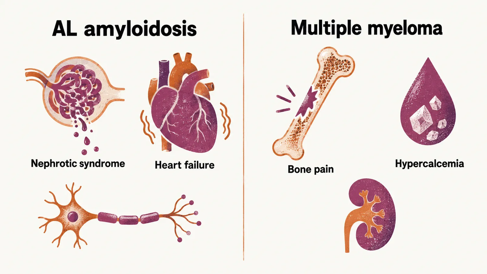
AL amyloidosis is the form of amyloidosis that is built from immunoglobulin light chains, and it is the amyloidosis that belongs to the plasma cell disorders.
Amyloidosis
Amyloidosis is a pathological condition in which a variety of proteins is deposited in the extracellular matrix of tissues. These proteins are normally soluble, while in the pathological condition of amyloidosis they undergo conformational changes, mainly the formation of anti-parallel beta-pleated formations, that make them insoluble. The misfolded protein then stacks into fibrils that resist clearance and slowly crowd the tissue.
In the case that amyloidosis is suspected, anti-parallel beta-pleated conformational protein has to be found in the extracellular matrix of the tissue. One of the easy diagnostic ways for amyloidosis is the use of Congo Red staining for the tissue: if there is anti-parallel beta-pleated amorphous material there, it is seen by polarised light as apple-green birefringence.
Different proteins can precipitate in the extracellular matrix of tissues and make amyloidosis; in the entity of plasma cell dyscrasia, only one specific form of amyloidosis matters, which is AL amyloidosis. AL amyloidosis is a monoclonal gammopathy that makes anti-parallel beta-pleated amorphous proteins which are specifically made out of light chain immunoglobulins.
Findings in AL amyloidosis
In the case of AL amyloidosis there is:
- an M spike in the serum of the patient
- bone marrow infiltrate by plasma cells
- amyloid amorphous material in the extracellular matrix of the kidney
- renal failure, which is totally different from MM’s renal failure: there is no cast nephropathy, and instead of tubulopathy there is glomerulopathy, which gives nephrotic syndrome
- hepatomegaly
- carpal tunnel syndrome
- otherwise unexplained heart failure
The organ pattern follows where the amyloid is laid down. Amyloid in the glomerulus leaks protein, so the patient presents with nephrotic syndrome rather than the tubular injury of myeloma; amyloid in the heart stiffens it, so the failure appears without a clear cardiac cause.
How AL amyloidosis differs from multiple myeloma
From the molecular oncogenesis point of view, AL amyloidosis and multiple myeloma are very similar to one another, because they are both made by the clonal malignancy of plasma cells. The difference between AL amyloidosis and MM is clinical: in amyloidosis there is heart failure and peripheral neuropathy, while in MM there is hypercalcemia, bone pain, renal failure and others.
If just one biochemical difference had to be found between AL amyloidosis and MM, it would be that in amyloidosis there is precipitation of antibodies, while in MM there is circulation of antibodies, which leads to hyperviscosity.