
The proteins that plasma cell disorders overproduce are immunoglobulins, and a monoclonal immunoglobulin — the M protein — is how these disorders are found in the blood and urine. A clone is a population of cells descended from one cell, so a monoclonal protein is one immunoglobulin made by one clone. To recognise it, it helps first to know how immunoglobulins are built and how they can differ from one another.
The structural categories of immunoglobulins
An immunoglobulin is built from two heavy chains and two light chains, and its variation can be described at three levels: isotype, allotype and idiotype. They run from the broadest difference, between classes, to the narrowest, the identity of a single clone.
- Isotype. Isotype differences are the differences in the constant heavy and light chains, so they define the classes of immunoglobulin rather than the individual molecule. The heavy-chain isotypes are IgA, IgG, IgM and others; the light-chain isotypes are the kappa and lambda chains. Every individual of a species carries the same isotypes, while the constant heavy chain differs between species.
- Allotype. Allotypes are small, regular differences in the amino acid sequence between individuals of the same species, produced by allelic differences in their immunoglobulin genes. They are the reason one person’s IgG can be recognised as foreign to another.
- Idiotype. The idiotype lies in the variable regions of the heavy and light chains, which are created by gene recombination when the B cell is formed. Because the variable region is the antigen-binding site, the idiotype is different for every clone of B cells or T cells, and it is the part of the molecule that marks a cell as belonging to one clone.
Electrophoresis and the M spike
By electrophoresis the proteins of the blood are separated into bands, and the way the gamma region looks is what distinguishes a normal serum from a clonal one. In the normal condition the gamma region contains many different immunoglobulins made by many different clones, so it appears as a broad peak, or hill. In a plasma cell disorder a single clone makes one immunoglobulin, so the same region becomes a narrow, sharp peak called the M spike. This change from a hill to a sharp peak is why these disorders are called gammopathies — the gamma region of the electrophoresis changes from a hill to a peak.
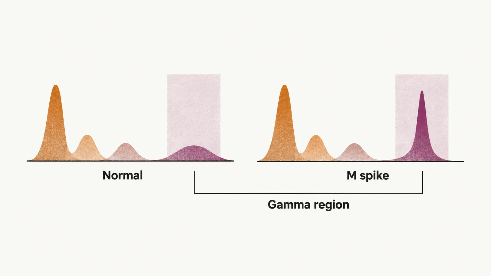
Serum protein electrophoresis, or SPEP, is a semi-quantitative measurement: on the strip, the more intense the band, the higher the amplitude of the spike. Electrophoresis is used for quantitative measurement. It can detect a monoclonal protein once there is at least about 0.5 g/dL; below that amount the M spike is not seen, which is one reason a normal electrophoresis does not exclude a plasma cell disorder.
To identify which isotype the protein belongs to, or to separate heavy from light chain, immunofixation is used, and it is a qualitative test.
What an M component can be
Once a monoclonal protein is detected, the next question is what it consists of. The nature of M components in plasma cell disorders can be:
- an intact antibody
- an altered antibody
- a fragmented antibody
- isolated light or heavy chains
There is no M spike in some forms of plasma cell disorders, such as in two-thirds of solitary bone plasmocytoma or extramedullary plasmocytoma.
Light chains and the kidney
Isolated light chains are also produced by normal plasma cells. In most plasma cells, light chains are produced in a slightly higher proportion than heavy chains, so some light chain is always left over after the molecules are assembled. This excess light chain is secreted from the cell and cleared from the body with the help of the kidney. The normal amount of light chain excreted is small, on the order of a few milligrams per day (less than 10 mg/day).
An M component is not specific for plasma cell disorders
The M component marks a clonal expansion of B cells or plasma cells, but not necessarily a plasma cell disorder. It can also be seen in:
- hematological cancers: CLL (chronic lymphocytic leukemia), CML (chronic myeloid leukemia), and B and T cell lymphomas
- non-hematological cancers: breast cancer, colon cancer
- non-neoplastic conditions: cirrhosis, sarcoidosis, Gaucher’s disease, pyoderma gangrenosum
- autoimmune conditions: RA (rheumatoid arthritis), MG (myasthenia gravis), cold agglutinin disease
- transplant patients
- some rare skin diseases: lichen myxedematosus, necrobiotic xanthogranuloma
It would be fair to say that 95% of monoclonal gammopathies are disorders related to plasma cells; the reason that not all of them are is Waldenström macroglobulinemia, also called lymphoplasmacytoid lymphoma, in which the clone is a lymphoid cell rather than a plasma cell.