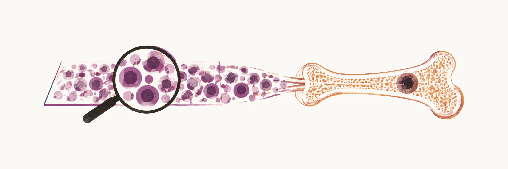
The three components of the diagnosis
The diagnosis of multiple myeloma needs three components:
- marrow plasmacytosis, which is defined as more than 10% plasma cells
- a serum and/or urine M component
- the presence of at least one myeloma defining event
Bone marrow plasma cells are CD138+ and either monoclonal kappa or lambda positive: CD138 is a surface marker of plasma cells, and monoclonality is shown by the clone making only one of the two light-chain types.
There are two variants of multiple myeloma:
- solitary bone plasmocytoma: a single lytic bone lesion without marrow plasmacytosis
- solitary extramedullary plasmocytoma: it usually involves the submucosal lymphoid tissues of the nasopharynx or the paranasal sinuses
The investigations below follow the three components: the bone marrow biopsy shows the plasma cells, the laboratory tests show the M component, and imaging looks for the bone lesions that count as myeloma defining events.
Bone marrow biopsy
A bone marrow biopsy is the investigation that establishes marrow plasmacytosis. Several tests are performed on the bone marrow cells:
- checking the morphology of the cells to find plasma cells
- checking the surface markers by flow cytometry
- checking the karyotype by FISH
- immunohistochemistry, in which Ki-67 is used as a detector for the S-phase protein of proliferative cells
Laboratory investigation
The laboratory investigation looks for the M component in serum and urine. Because in some patients the M component is made of light chains only, several complementary tests are used.
Serum protein electrophoresis, or SPEP (semi-quantitative test)
The serum M component is made up of IgG in 53% of cases, IgA in 25%, IgD in 1% and just light chains in 20% of cases. In the case that the light chain is the only protein that is made by the MM cells, no M spike is seen on the SPEP, so an immunofixation test is needed. 97% of cases of MM are secretory forms, while in 3% of cases there is no secretion of Ig.
Measurement of the serum immunoglobulin level
The serum immunoglobulin level is also measured.
Serum immunofixation, or IF (qualitative test)
Immunofixation is the test to do when the aim is to be sure that the gamma peak seen on the electrophoresis is related to a monoclonal antibody. The workup of IF uses staining specific for the immunoglobulin, like IgG or IgM, to see which immunoglobulin isotype the peak in the gamma region is related to; mostly five different specific stains are used, for IgG, IgM, IgA, kappa light chain and lambda light chain. In the case of light-chain-only MM, in which no M spike can be detected by the SPEP, immunofixation is one way to be sure about the presence of this form of MM.
Serum free light chain
Serum free light chain is mandatory in this setting, because in the 20% of cases of secretory MM in which the immunoglobulins are only light chains there is no spike on the SPEP, so this test is used to be sure about the presence of light chains in the serum.
24-hour urine for light chains
The 24-hour urine is used for measurement of light chain, both quantitative and by electrophoresis. Dipsticks for light chains do not work. Fewer than 1% of cases do not have any M components, which could be due to a high catabolism rate by the kidney in these patients that makes the M component invisible; in these patients the serum M component for light chain can nowadays be detected.
The peripheral blood smear
In the peripheral blood smear of patients with MM there is no specific marker related to plasma cells, but casts of erythrocytes can be seen, made by the attachment of the erythrocytes by excess antibodies in the serum; this is called Rouleaux formation. Rouleaux formation is not specific for MM: it can be seen in any condition with hypergammaglobulinemia, like infectious mononucleosis or liver cirrhosis.
Radiological investigation
The radiological investigation for patients who are suspected of having MM, or any form of plasmocytoma, is as follows:
- The very first test that has to be performed is a CT of the whole body. It is very useful for the detection of fractures in the bones, like a spine fracture, which can happen in the case of MM, because CT shows the mineralised bone well.
- If the CT is negative in these suspected patients, the next step is a whole body MRI. MRI is more sensitive than CT for the detection of areas in the bone that are full of plasma cells in the case of MM, because MRI reads the marrow itself. CT is more useful for the detection of damage to the bones, while MRI shows the presence of lytic activity of plasma cells in the bone.
Distinguishing myeloma from its precursor states
The most important differential diagnosis of patients with multiple myeloma is related to monoclonal gammopathy of undetermined significance (MGUS) and smoldering multiple myeloma (SMM), the precursor states that share the same M protein without the end-organ damage. MGUS is very common in comparison to MM: it can be found in 3.2% of the population aged 50 or older and 5.3% of the population aged 70 or older.