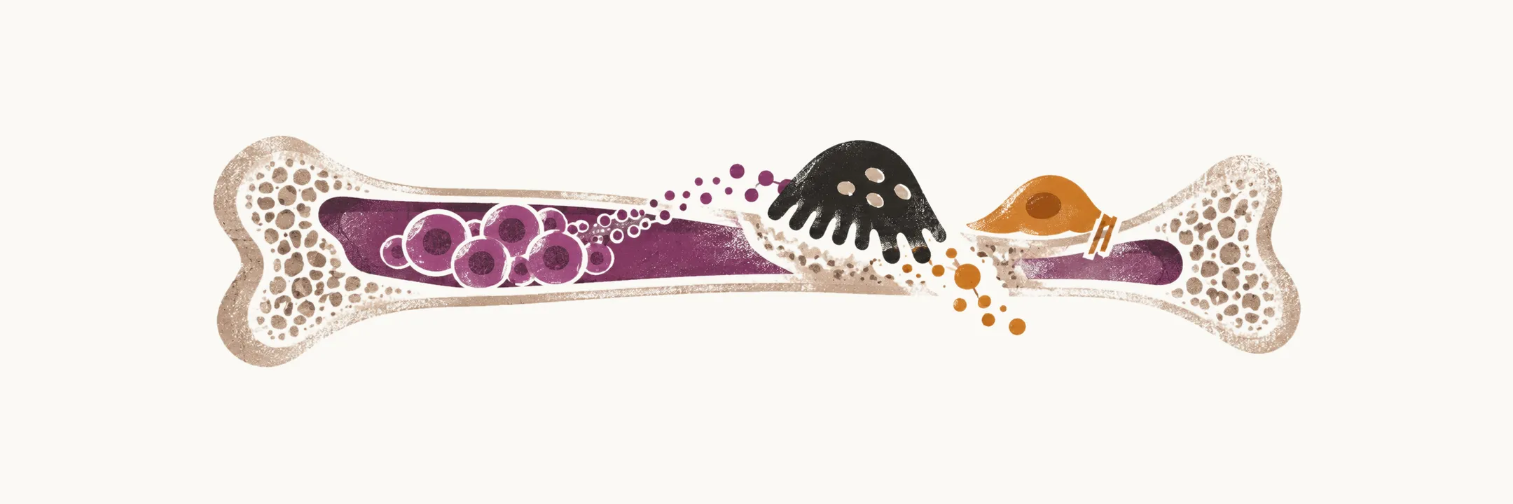
Multiple myeloma damages the body through the myeloma cells themselves and through what those cells release. The myeloma cells do not grow in isolation: they depend on the surrounding bone marrow, and that dependence is the first step in understanding the disease.
How myeloma cells live in the bone marrow
MM cells are attached to the bone marrow stromal cells, or BMSCs, and to the extracellular matrix, or ECM, with the help of cell-surface adhesion molecules. The attachment is not merely a way of holding the cell in place: it switches on the signals the myeloma cell needs to survive, and the result is survival, growth, resistance to drugs and migration of the cells to the bone marrow. Each of these runs through a particular cascade — growth mainly through the ras/raf/MAPK cascade, drug resistance mainly through the PI3K/Akt cascade, and migration mainly through the PKC cascade.
These effects are due to either direct cell-BMSC interaction or the effects of cytokines. In direct interaction, the endothelial cells and the osteoclasts are the main cells that support MM cells, and pDCs (plasmacytoid dendritic cells), MDSCs (myeloid-derived suppressor cells) and Th17 cells also take part. The cytokines involved are VEGF, IL-6, IGF and stromal-derived growth factor, or SDF-1α.
Among cytokines, IL-6 has an evident role in driving myeloma proliferation: it is released by the bone marrow stromal cells that surround the tumour and acts on the myeloma cell to keep it dividing and to protect it from apoptosis.
Clinical features
The clinical manifestation of MM is due to two main patterns made by the MM cells: the uncontrolled formation of antibodies and the infiltration of malignant cells into the bone marrow. The tumour and its products, beside the host response, lead to multiple organ dysfunction or failure and some main symptoms.
Bone fracture or pain
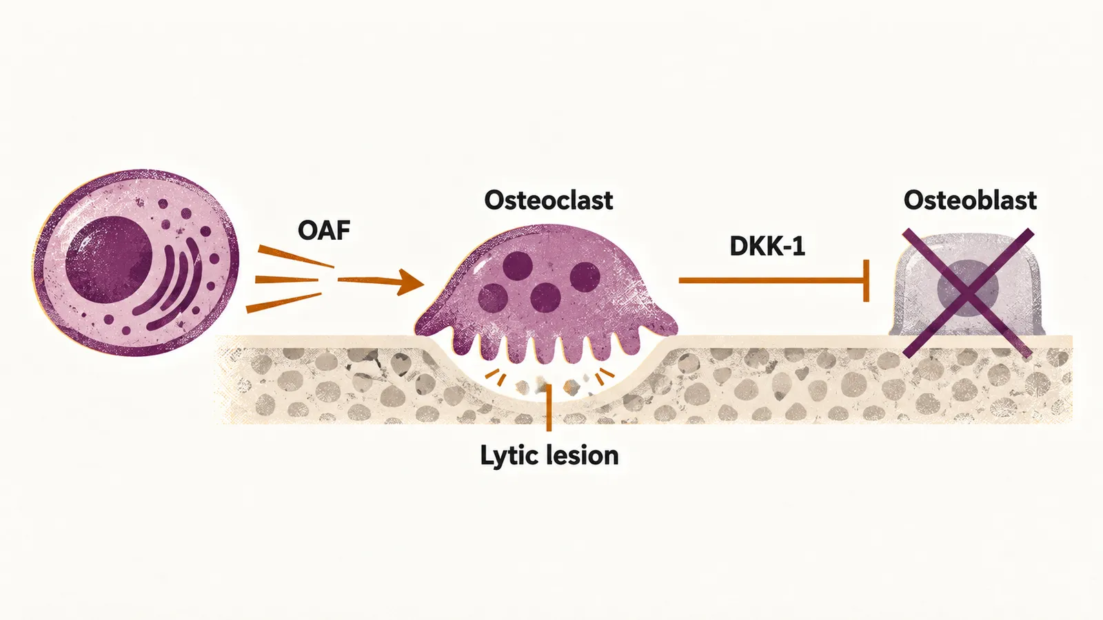
Bone pain is the most common symptom in patients with MM, and about 70% of cases are affected by it. Bone is normally kept in balance by osteoclasts, which resorb it, and osteoblasts, which form it. The bone lesions in MM are due to the proliferation of tumour cells, the activation of osteoclast cells and the suppression of osteoblasts, and these changes finally lead to the formation of bone lesions.
The activation of osteoclast cells is mediated by osteoclast activating factor (OAF), which is made by the myeloma cells after the triggering of several cytokines like VEGF, TNF-α, IL-1, RANK ligand and some others. The inhibition of the activity of osteoblast cells is mediated by Dickkopf-1 (DKK-1), which is also made by the myeloma cells. Because the normal balance between bone formation and bone resorption is lost in both directions at once, the lesions are lytic and do not heal.
Hypercalcemia
Hypercalcemia is mainly due to the activation of osteoclast cells, which leads to the release of calcium ions from the bone matrix into the blood. The raised calcium explains the thirst, polyuria, constipation and confusion that can accompany it.
Susceptibility to infection
After bone pain and fracture, the most common problem in MM patients is susceptibility to bacterial infections like S. pneumoniae, S. aureus and K. pneumoniae, and pyelonephritis, mainly by E. coli. The main cause of these infections in patients with MM is diffuse hypogammaglobulinemia, if the M component is excluded, meaning a low level of the normal antibodies apart from the M protein, which is due to the destruction of normal antibodies and the reduction of production of normal antibodies.
Beside the fact that normal antibodies are decreased in MM patients, some other factors also contribute to the susceptibility to infections: a decreased Th1 response, an increased Th17 response, an aberrant Treg response, a low granulocyte lysozyme content and a low migration rate for granulocytes.
Renal failure
Renal failure is present in more than 25% of cases of MM, and it can happen for several different reasons: hypercalcemia, which is the most common cause of renal failure in MM patients; the deposit of amyloid; hyperuricemia; recurrent infections; recurrent use of NSAIDs for bone pain; iodinated contrast agents; and bisphosphonate use. Because of the hypercalcemia that MM patients have, osmotic diuresis can also be seen, which can lead to dehydration and finally pre-renal insufficiency, beside all the other forms of renal injury described here.
The kidney tubules and glomeruli can be damaged in different ways. Tubular damage, or tubulopathy of the distal tubules due to the deposition of light chains, is almost always present. The earliest manifestation of tubular damage in adults is Fanconi’s syndrome, which is characterised by the loss of amino acids, the loss of glucose, the inability of the kidney to acidify the urine and the inability of the kidney to concentrate the urine; the proteinuria here is not accompanied by hypertension. When MM also damages the glomeruli, it leads to non-selective proteinuria with the presence of albumin and hypertension in the patient. Lambda light chains are more potent than kappa light chains in the formation of renal injury and amyloidosis in the kidney.
Anemia
Anemia is the most common sign. Normocytic and normochromic anemia occurs in more than 80% of MM patients, and it is related mostly to the replacement of normal marrow cells with tumour cells.
Clotting problems
Clotting challenges in MM patients happen mostly because of the covering of normal platelets by antibodies that prevent them from functioning normally. Beside the attachment of antibodies to the platelet, the M component also attaches to clotting factors such as factors I, II, V, VII and VIII, and there are amyloid damages to the endothelial cells.
Neurologic symptoms
A vertebral lesion can press on the spinal cord or on a nerve root, producing back pain, pain radiating along a limb, weakness or loss of sensation, and compression of the cord is a neurological emergency. Peripheral neuropathy also occurs, most often when amyloid is deposited in the nerves.
Hyperviscosity of blood
The different isotypes of immunoglobulin have different effects on the viscosity of blood: IgM has more effect on the increase of viscosity in comparison to the IgG, because IgM circulates as a large pentamer that stays in the plasma rather than distributing into the tissues.