Diagnosing dermatomyositis starts with suspicion, because the rash plus proximal weakness identifies the disease before any laboratory result returns. The workup then serves three purposes at once: confirming the diagnosis, measuring activity for treatment decisions, and screening for the malignancy that adult-onset disease can signal.
The rash leads
The heliotrope discoloration with periorbital swelling distinguishes dermatomyositis from every other myopathy, which is why patients often arrive via dermatology and why amyopathic disease can be diagnosed on cutaneous grounds alone. In early or mild disease the rash may be the only abnormality. A classic clinical picture outweighs an inconclusive biopsy, a hierarchy worth remembering when the two disagree.
Blood markers of fibre injury
Active inflammation spills intracellular enzymes into blood. Creatine kinase (CK) is the most sensitive and practical, tracking activity closely enough to follow treatment: falling values accompany response, rising values warn of relapse. Aldolase adds value when CK is normal yet suspicion runs high, while myoglobin and lactate dehydrogenase corroborate ongoing injury without the same specificity.
Autoantibodies and subsets
Myositis-specific antibodies, autoantibodies directed against the body’s own proteins and characteristic of myositis, appear in roughly a third of dermatomyositis and polymyositis patients and carry prognostic weight. Each one points to a different clinical picture:
- Anti-Mi-2 is closely specific to dermatomyositis with classic skin disease and predicts a good steroid response.
- Anti-Jo-1, the commonest antibody overall, defines the anti-synthetase syndrome of myositis with interstitial lung disease, nonerosive arthritis, Raynaud phenomenon, mechanic’s hands, and fever, where lung disease may dominate management and threaten life.
- Anti-MDA5 identifies a phenotype in which the skin may be severely affected, or the weakness minimal, while interstitial lung disease can progress rapidly, so a positive result prompts urgent assessment of the lungs; it is more common in East Asian populations.
- Anti-TIF1-γ is strongly associated with cancer in adult-onset disease and supports thorough malignancy screening.
- Extractable nuclear antigen antibodies, including anti-Scl-70, anti-SSa and SSb, anti-Sm, and anti-RNP, mark overlap with systemic sclerosis, Sjogren disease, lupus, or mixed connective tissue disease.
Because the antibody profile predicts which organs are involved and how the disease behaves better than the clinical label alone, serotype-based classification, formalised in the 2017 EULAR/ACR criteria, increasingly guides expectations.
The biopsy hallmark and its limits
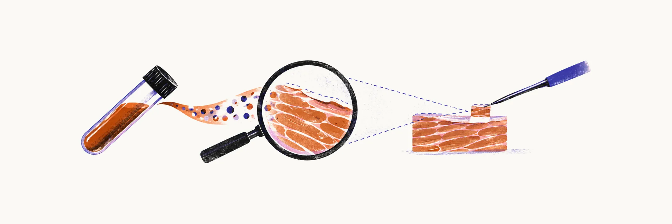
Perifascicular atrophy, wasting concentrated at fascicle edges with relative central sparing, is considered pathognomonic, meaning distinctive enough to establish the diagnosis. Supporting features include reduced capillary density and membrane attack complex deposition on surviving capillaries. Two caveats protect against overreliance. Inflammatory infiltrates may be sparse or absent, unlike polymyositis where endomysial invasion is required, and early disease may not yet show the atrophy pattern. A negative biopsy therefore never excludes dermatomyositis when rash and weakness fit; serology and observation, sometimes with repeat sampling, carry the diagnosis.
Cancer screening
Adult-onset disease obliges age-appropriate malignancy screening at diagnosis: thorough history and examination including breast, pelvic, and rectal assessment, blood work, chest imaging with CT preferred for occult lung lesions, abdominal and pelvic imaging for ovarian, pancreatic, and gastrointestinal primaries, colonoscopy by age guideline, and mammography. PET-CT suits high-suspicion or atypical cases. Because paraneoplastic disease, disease driven by the remote effects of a tumour, can precede detectable tumour by months or years, screening repeats when disease resists adequate immunosuppression or returns after remission.
The sequence in practice
Taken in order, the steps move from suspicion to confirmation to screening.
| Step | Test | What it establishes |
|---|---|---|
| 1 | Examination for rash with proximal weakness | Diagnostic suspicion and separation from polymyositis and inclusion body myositis |
| 2 | Serum CK, aldolase, myoglobin, LDH | Active fibre injury and a baseline for monitoring |
| 3 | Antibody panel including anti-Mi-2, anti-Jo-1, anti-MDA5, anti-TIF1-γ, ANA (antinuclear antibodies), and overlap antibodies | Support for dermatomyositis, anti-synthetase, anti-MDA5, cancer-associated, and overlap subsets |
| 4 | Muscle biopsy when the picture is unclear | Perifascicular atrophy confirms; a negative result does not exclude |
| 5 | Age-appropriate cancer screening | Detection of paraneoplastic malignancy in adult onset |