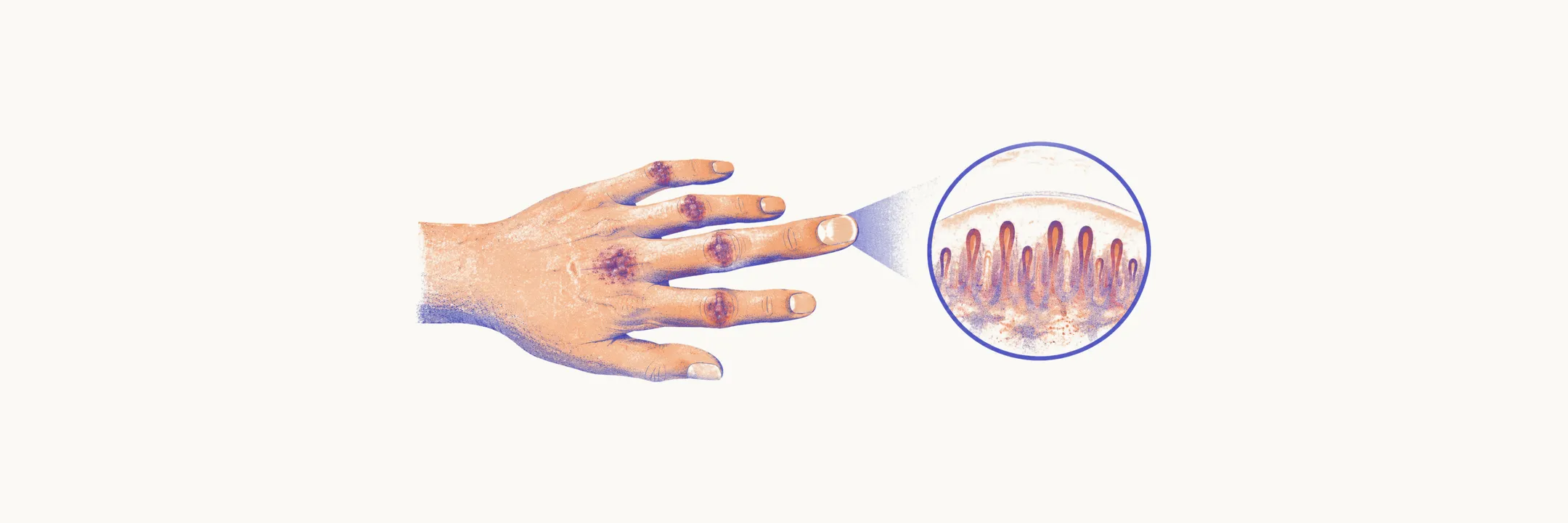
Skin disease is the defining feature of dermatomyositis and affects the large majority of patients. It often appears months before any weakness, and in some patients it is the only manifestation of the disease. The pattern matters for two reasons: the rash alone can establish the diagnosis, and particular cutaneous findings point to particular antibody subsets and to organs beyond the skin.
The characteristic signs
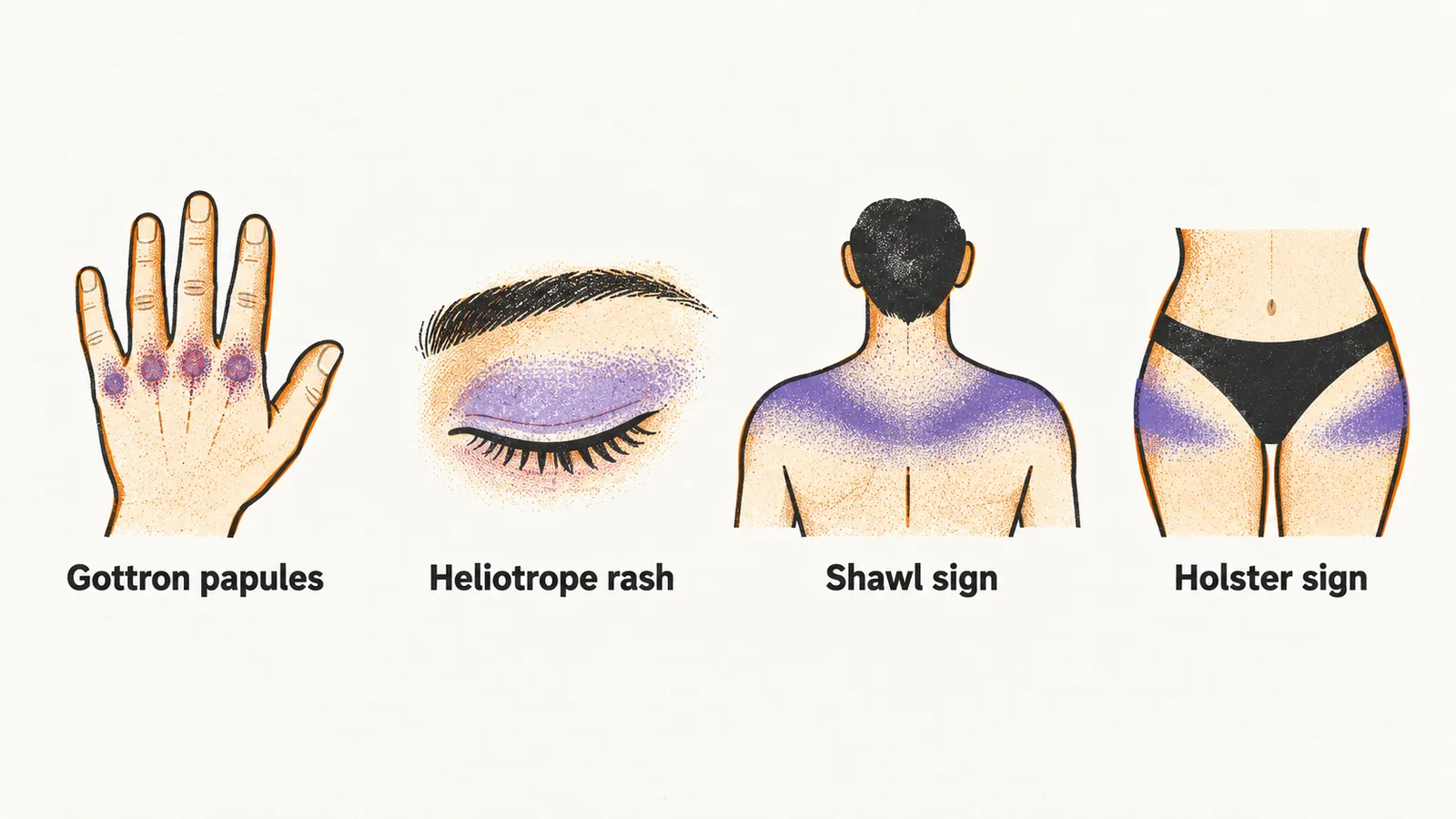
Several signs have names of their own. They are easiest to learn by the part of the body they mark: the hands, the face, and the neck, trunk, and thighs.
The hands
Gottron papules are violaceous (purple-tinged), slightly raised lesions on an erythematous (red) base over the extensor surfaces of the small joints of the hands, classically the metacarpophalangeal and interphalangeal joints. Flat erythematous patches in the same territory, sometimes scaling and sometimes extending to the elbows and knees, are called Gottron sign. Mechanic’s hands describes hyperkeratotic (thickened), fissured skin along the lateral margins of the fingers and fingertips, and it points toward the anti-synthetase overlap in which interstitial lung disease can dominate.
The face
The heliotrope rash is a violaceous discoloration of the upper eyelids, often with periorbital swelling, and it can wax and wane with disease activity. Lupus can mimic the eyelid change but typically spares the upper lids, which is a useful distinction at the bedside.
The neck, trunk, and thighs
The shawl sign is an erythematous eruption across the posterior neck, shoulders, and upper back, and the V sign is its equivalent on the anterior chest. A poikiloderma-like change (skin showing pigment change, atrophy, and telangiectasia together) over the lateral thighs and hips is known as the holster sign.
The rash prefers sun-exposed skin, characteristically the face, posterior neck, upper back, and the V of the chest, while covered skin tends to be spared. Photosensitivity is a recognised feature of the disease and is especially marked in the anti-TIF1-γ subset.
Nailfolds
Nailfold change is common and easy to look for: periungual (around the nail) telangiectasia, meaning dilated small surface vessels, overgrowth or dystrophy of the cuticle, and punctate haemorrhages. On nailfold capillaroscopy, examination of the nailfold capillaries under magnification, the capillary loops are dilated and tortuous and their density is reduced, which is the same microvascular injury seen in muscle, and higher capillaroscopy scores track with greater disease activity.
Ulceration and palmar papules
Two findings belong to a more severe cutaneous phenotype. Painful papules on the palms, particularly over the joints, are called inverse Gottron papules, and skin ulcers may form over Gottron lesions, on the digital pulp, or around the nailfolds. Both are associated with the anti-MDA5 antibody and with interstitial lung disease, an inflammatory and scarring disease of the lung tissue.
Calcinosis
Calcinosis is the deposition of calcium in skin and subcutaneous tissue. It develops in a substantial proportion of children and adolescents, classically around 2 in 5, while remaining rare in adults. The deposits form hard nodular masses, most often over pressure points such as the elbows, knees, and buttocks, and are visible on imaging. They can cause pain, contracture, and functional loss, and their frequency tracks with delayed or inadequate treatment, which is one more reason early therapy matters. Calcinosis with myositis in a child points to juvenile dermatomyositis first; in an adult it suggests chronic or undertreated disease.
How the skin behaves over time
The skin does not always follow the muscle. The rash can persist or flare after strength and creatine kinase (CK, a muscle enzyme that rises with fibre injury) have returned to normal, and it may leave permanent pigmentary change, atrophy, or telangiectasia. Cutaneous disease therefore has its own course and can remain active after the muscle disease is controlled.
Skin disease without weakness
A minority of patients have the typical rash with no clinical muscle weakness, which is called amyopathic dermatomyositis. When laboratory or electrical testing reveals subtle muscle involvement that the patient does not feel, the term hypomyopathic is used instead. Some of these patients develop weakness months or years later and some remain purely cutaneous, but the diagnosis stands on skin grounds with skin biopsy support. Skin-only disease is not necessarily mild: interstitial lung disease and, in adults, malignancy are recognised in this group.