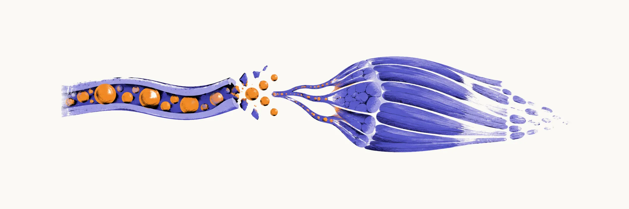
Dermatomyositis is an immune-mediated disease of skin and skeletal muscle in which the main target is not the muscle fibre (the muscle cell) itself but the small blood vessels that supply it. That shift of target explains the two features that define the disease, the rash and the proximal weakness, and it also explains the pattern a muscle biopsy shows.
The capillary as the first target
The injury begins in the microvasculature. Complement, a set of plasma proteins that mark and puncture target cells, is activated in muscle, and its final product, the membrane attack complex (C5b-9), is deposited on the walls of muscle capillaries. Capillaries are destroyed, capillary density in muscle falls, and the surviving vessels carry the same deposits. Because the same small-vessel injury occurs in skin, the rash and the muscle disease belong to one process rather than two.
From capillary loss to perifascicular atrophy
A muscle fascicle is a bundle of muscle fibres. In dermatomyositis the fibres at the outer edge of each fascicle shrink while fibres in the middle of the bundle are relatively spared, a pattern called perifascicular atrophy. The long-standing explanation is ischaemia: perifascicular fibres sit at the end of the capillary supply, so they are the first to suffer once capillaries are lost.
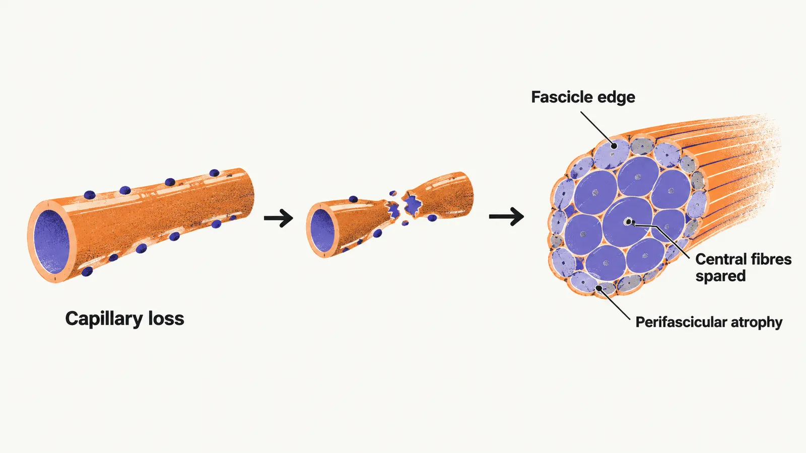
Two observations have made that explanation less secure. The skin lesion follows the same border pattern, injuring the basal cells of the epidermis where it meets the dermis, and the epidermis has no blood vessels at all, so ischaemia cannot account for it. And type 1 interferon-inducible proteins such as MxA and ISG15 are markedly overproduced in dermatomyositis muscle and skin, more so than in the other muscle diseases studied. Interferon signalling may therefore injure perifascicular fibres and basal keratinocytes directly, with capillary damage and perifascicular atrophy as two consequences of one process rather than one causing the other.
Type 1 interferon signalling and the antibody link
Type 1 interferons are innate immune signalling molecules, the group that includes interferon alpha and interferon beta. In dermatomyositis, interferon-inducible genes and proteins are increased in both muscle and skin, and the endothelial cells of affected muscle contain tubuloreticular inclusions, which are structural markers of interferon exposure. The pathway also connects the disease to one of its antibody subsets. MDA5 (melanoma differentiation-associated gene 5) is a cytosolic sensor of viral RNA whose activation induces type 1 interferon, and antibodies against MDA5 mark a form of dermatomyositis in which weakness may be minimal while vascular injury and fibrosis concentrate in the lungs, producing interstitial lung disease that can progress rapidly. Anti-MDA5 disease is more common in East Asian populations than in European ones.
How the mechanism differs from polymyositis and inclusion body myositis
Setting this mechanism beside its two closest relatives shows what is specific to dermatomyositis. Polymyositis injures the fibre directly. Cytotoxic CD8-positive T cells, immune cells that kill their targets, invade muscle fibres that are not yet necrotic (dead), the fibres display MHC class I molecules on their surface, and the infiltrate lies within the fascicle, among the fibres (endomysial), rather than at its edge. That is why polymyositis produces neither a rash nor a perifascicular pattern. Inclusion body myositis combines an inflammatory infiltrate with protein aggregation, visible as rimmed vacuoles on biopsy, together with a degenerative component; the combination is why it does not respond to immunosuppression, unlike dermatomyositis and polymyositis.
Why the timing of treatment matters
Capillary loss and perifascicular atrophy are not fully reversible. Treatment therefore rescues fibres that have not yet been wasted rather than repairing what is already gone, so how much strength returns depends on how much atrophy had developed before the inflammation was controlled.