Fever with a rash is one of the most common presentations in paediatric practice, and the six classic exanthems look broadly similar at a glance: an irritable, febrile child covered in red spots. They are separated by mechanism rather than by appearance alone, so the diagnostic approach is to narrow the field with history and then read the rash against what remains. Four axes carry almost all of the discriminating information.
The four axes
| Axis | The question it answers | Where the answer comes from |
|---|---|---|
| Exposure | Is there a known contact, a nursery or school outbreak, recent travel, or vaccination status? | History |
| Age | Which of the six occurs at this age? | History |
| Season | Does the time of year fit? | History |
| Rash characteristics | Morphology, distribution, relationship to fever, and any enanthem (the same kind of eruption on a mucous membrane) | Examination |
Age and rash characteristics do most of the work. Exposure and season narrow the differential, but neither confirms or excludes anything on its own.
Age
Several of the six have a characteristic age band, so age narrows the field before the child is examined.
| Age band | Most likely classic exanthem |
|---|---|
| Under 3 months | Rarely any of the six — maternal antibody is still protective |
| 6 months to 2 years | Roseola infantum (human herpesvirus 6, HHV-6) |
| School-age children | Scarlet fever (group A Streptococcus), fifth disease (parvovirus B19) |
| Any age, unvaccinated | Measles, varicella, rubella |
A child under 3 months with a febrile rash is a different problem: at that age a serious bacterial illness has to be considered before any diagnosis of a viral exanthem is made.
Season
Time of year adds less than age does. Parvovirus B19 infection is more frequent in late winter, spring and early summer. Respiratory-transmitted infections, scarlet fever among them, cluster when children are crowded together indoors. Measles and roseola, by contrast, occur year-round in susceptible populations. That is why season is the weakest of the four axes: it can narrow the differential, but it should never settle the diagnosis by itself.
Rash characteristics
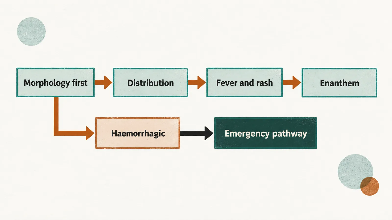
The examination reads the rash in four steps: the type of lesion, where it appears and how it spreads, when it appears relative to the fever, and whether there is an enanthem in the mouth. Each step removes some of the six.
Morphology first
Maculopapular (flat macules and raised papules) or vesicular (small fluid-filled blisters)? If the lesions are vesicles — clear fluid-filled, and above all present in crops at different stages of evolution — varicella is the working diagnosis. If the lesions are macules and papules, the rest of the assessment decides which of the other five it is. A haemorrhagic rash, made of bleeding into the skin, takes the child out of this framework altogether and into the emergency pathway.
Distribution
Three terms describe the direction of spread: cranio-caudal means from the head downwards, centrifugal means from the trunk outwards to the face and limbs, and centripetal means concentrated on the trunk more than on the limbs.
| Pattern | Suggests |
|---|---|
| Head then trunk then limbs (cranio-caudal) | Measles |
| Trunk, spreading outwards, accentuated in the flexures (skin creases); spares the perioral area (around the mouth) | Scarlet fever |
| Trunk, then face and limbs (centrifugal) | Roseola |
| Slapped cheeks, then lacy reticulated (net-like) rash on the limbs | Fifth disease |
| Trunk-dominant, centripetal, lesions at several stages | Varicella |
| Face first, fading within about 3 days | Rubella |
The relationship between rash and fever
| Pattern | Suggests |
|---|---|
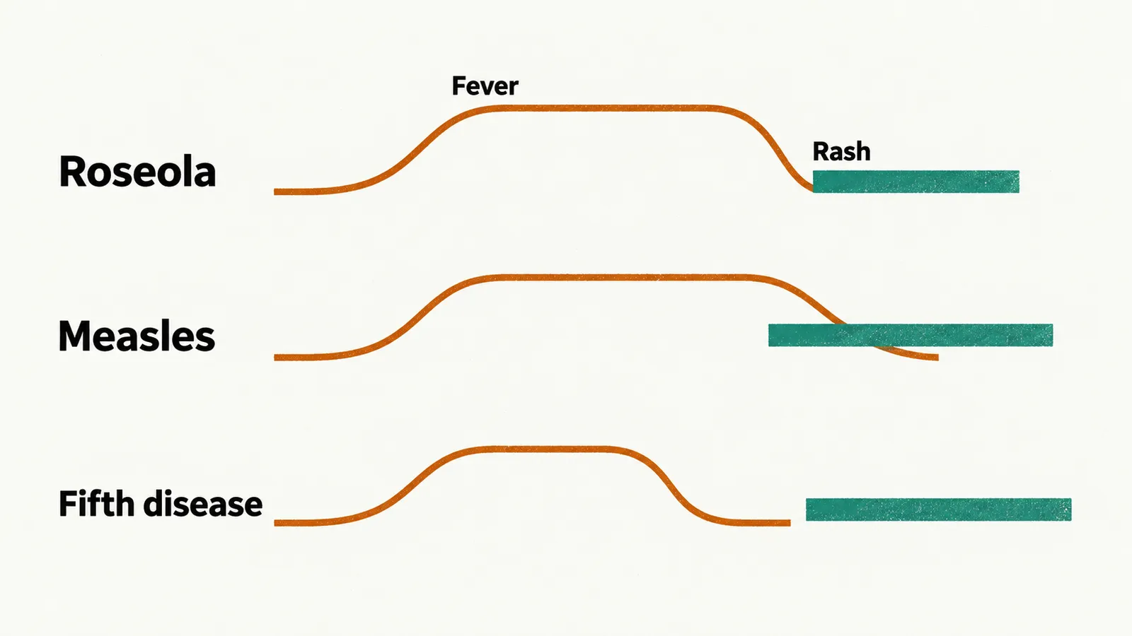
| Fever for several days, then rash as the fever breaks | Roseola |
| Fever, then rash while the fever continues | Measles, scarlet fever, varicella |
| Rash and fever together, without a preceding febrile phase | Most other exanthems |
| No fever by the time the rash appears | Fifth disease (the fever belongs to the earlier viraemic phase, when virus is circulating in the blood), rubella |
Enanthem, when present, is the most specific finding
| Enanthem | Disease |
|---|---|
| Koplik spots (small white lesions on the buccal mucosa) | Measles |
| Strawberry tongue (white coating with red papillae, later a red tongue with prominent papillae) | Scarlet fever |
| Palatal petechiae or small red spots | Rubella |
| Palatal vesicles | Varicella |
| None | Roseola, fifth disease |
Koplik spots appear during the prodrome (the symptomatic phase before the rash) and disappear as the rash appears, so they are visible only in a narrow window.
The six diseases side by side
Once the rash has been read and one diagnosis is likely, the practical questions change: how long the child is contagious, whether the diagnosis needs confirming, what treatment and prevention exist, and which complication to watch for. The table sets those rows beside the pathogen and the rash features above. In the confirmation row, IgM is the antibody class that marks a recent infection, and RT-PCR detects the virus’s genetic material in a swab or sample. In the complication row, subacute sclerosing panencephalitis is a late, fatal brain disease caused by persistent measles virus; a transient aplastic crisis is a temporary arrest of red cell production; and fetal hydrops is abnormal fluid accumulation in the fetus.
| Feature | Measles | Scarlet fever | Rubella | Fifth disease | Roseola | Varicella |
|---|---|---|---|---|---|---|
| Pathogen | Measles virus | Group A Streptococcus exotoxin | Rubivirus | Parvovirus B19 | HHV-6 | Varicella-zoster virus |
| Rash morphology | Maculopapular | Maculopapular, sandpaper-like | Maculopapular, faint | Maculopapular, slapped cheek then lacy | Maculopapular, pink | Vesicular, in crops |
| Distribution | Cranio-caudal | Trunk and flexures outwards | Face downwards, lasting about 3 days | Cheeks then limbs | Trunk then face and limbs | Centripetal, multiple stages |
| Enanthem | Koplik spots | Strawberry tongue | Palatal spots | None | None | Palatal vesicles |
| Fever and rash | Rash while fever continues | Rash while fever continues | Rash with mild or no fever | Child well, fever already resolved | Rash as fever breaks | Rash while fever continues |
| Contagious window | About 4 days before to 4 days after rash onset | During the acute illness; ends 24-48 hours after starting antibiotics | About 7 days before to 7 days after rash | Before the rash, during the viraemic phase | Viraemic phase, before the rash | 1-2 days before the rash until all lesions have crusted |
| Confirmation | Clinical suspicion; laboratory confirmation (IgM or RT-PCR) for every suspected case | Rapid antigen test, nucleic acid test or throat culture | Serology or PCR if needed; rash is not specific | Clinical; serology or PCR if needed | Clinical | Clinical |
| Treatment and prevention | Supportive, vitamin A; MMR | Antibiotics; no vaccine | Supportive; MMR | Supportive; no vaccine | Supportive; no vaccine | Antivirals in risk groups; varicella or MMRV vaccine |
| Main complication | Subacute sclerosing panencephalitis | Acute rheumatic fever, glomerulonephritis | Maternal-fetal infection (outside this family) | Transient aplastic crisis, fetal hydrops | Febrile seizures | Zoster, later in life |
When the pattern does not fit
Two situations should interrupt the framework immediately.
The first is a haemorrhagic rash: petechiae that do not blanch, purpura or ecchymoses in a febrile child. This is not the usual presentation of any of these six. Meningococcal and other invasive bacterial disease, severe measles, haemorrhagic fevers and coagulopathy belong at the top of the list, and the child needs urgent assessment and treatment rather than diagnostic refinement.
The second is a drug exposure in the preceding days or weeks. Many maculopapular eruptions are drug reactions, and a history of atopy (a tendency to allergic disease) with intense pruritus (itching) and rapid improvement when the trigger is withdrawn points that way.
Outside those two situations, the practical rule is that a well-appearing child with a characteristic rash and a matching age and exposure history does not need laboratory confirmation; a child who is unwell, or whose rash does not fit any pattern, does.