An exanthem is a widespread cutaneous eruption built from repetitive lesions: the same morphology repeated across the skin rather than a solitary or localised lesion. The word comes from Greek roots meaning “to flower outwards”, which describes the appearance reasonably well. The same phenomenon on a mucous membrane is an enanthem; both are common in the same illness, and an enanthem is often the earlier and more specific finding.
Exanthems have many causes, including drugs, toxins, microorganisms and autoimmune disease. The six classic childhood exanthems are the illnesses in which the rash is the clearest and most characteristic feature, rather than a subsidiary finding during some other infection.
Two mechanisms produce an exanthem
Direct damage to skin cells
The organism invades the epidermis or dermis (the outer and inner layers of the skin) and damages cells directly. This is the usual mechanism in the viral exanthems — measles, roseola, varicella — where viral replication in the skin or in dermal capillaries produces the visible lesion.
Damage to cutaneous vessels
Here the lesion comes from the small blood vessels of the skin rather than from the skin cells themselves. Damage to capillary walls produces erythematous (reddened) lesions, and when it is severe it produces petechiae and ecchymoses, small and larger areas of bleeding into the skin. Four sub-mechanisms are described:
| Sub-mechanism | Example |
|---|---|
| Toxin release | Scarlet fever (streptococcal pyrogenic exotoxin), staphylococcal toxin syndromes |
| Immune-complex deposition or cell-mediated response | Vasculitic exanthems |
| Direct damage by the microorganism | Meningococcal petechiae |
| Infiltration by phagocytes and lymphocytes | Rickettsial infections |
Fifth disease belongs in this second group as well: its rash is thought to be immune-mediated — immune-complex deposition rather than viral invasion of the skin.
The distinction matters because it explains where the organism actually is. In scarlet fever the streptococci are in the pharynx, not the skin: the rash is an immune reaction to a circulating exotoxin (a toxin the bacterium releases), appearing after the pharyngitis has been present for a day or more. Whether the rash appears depends on the immune response to the toxin rather than on the severity of the infection.
Primary lesions, and what each one implies
Whatever the mechanism, what the examiner sees is a set of individual lesions, and a rash is described by naming the lesion it is built from. The names are worth learning precisely, because each lesion type points to a process in the skin: flat lesions come from vasodilatation, raised ones from oedema or cellular infiltration, and bleeding into the skin from fragile capillaries, broken vessels or a coagulation abnormality.
| Primary lesion | Mechanism |
|---|---|
| Erythema | Vasodilatation |
| Macule (flat) | Vasodilatation |
| Patch | Vasodilatation or pigment accumulation |
| Papule (raised) | Oedema and cellular infiltration |
| Plaque | Confluence of papules |
| Nodule | Inflammatory infiltration of the deepest dermal layers |
| Vesicle | Cellular necrosis with intra- and intercellular oedema and acantholysis (loss of adhesion between epidermal cells) |
| Bulla | A larger vesicle |
| Pustule | Pus-filled lesion |
| Crust | Dried exudate from a previous lesion |
| Scale | Parakeratosis (retained nuclei in the outer horny layer) and dyskeratosis (abnormal keratinisation of individual cells) |
| Wheal | Oedema |
| Petechiae | Bleeding from dermal capillaries, from fragility or a coagulation abnormality |
| Ecchymosis | Subcutaneous blood collection from a broken vessel or coagulopathy |
Three of these names recur throughout the six classics: macules and papules, which make up five of them, and vesicles, which define varicella. Petechiae and ecchymoses are the opposite case: none of the six is primarily built from them, so they point away from the classics.
Classification by morphology
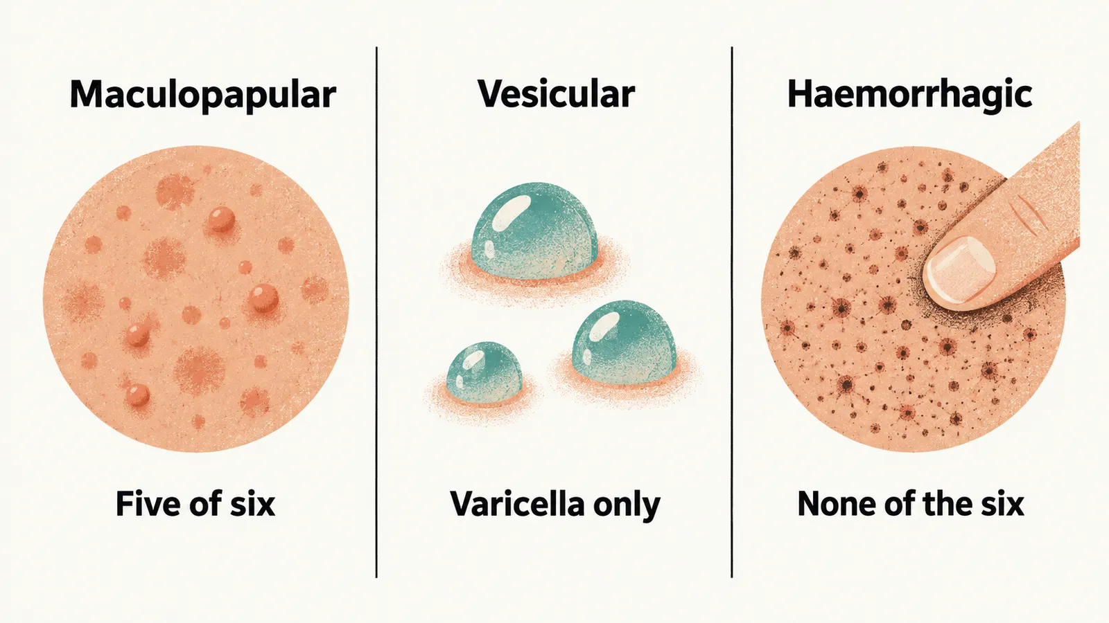
Grouping eruptions by their dominant lesion gives three morphological classes, and the six classics fall very unevenly among them.
Maculopapular
A maculopapular exanthem is made of macules and papules, flat and raised lesions. Five of the six classics belong here. Maculopapular exanthems also occur with many infections outside this group (dengue, Epstein-Barr virus, cytomegalovirus, HIV, rickettsial infections, typhoid, secondary syphilis, enteroviral illness), with drug reactions, and with fungal disease.
Vesicular
Varicella is the only classic in this group. Other vesicular eruptions include herpes zoster, herpes simplex and hand-foot-and-mouth disease.
Haemorrhagic
No classical childhood exanthem is primarily haemorrhagic. A haemorrhagic rash — petechiae that do not blanch, or ecchymoses — in a febrile child should be treated as an emergency and investigated for invasive bacterial infection (meningococcal disease above all), severe measles, a haemorrhagic fever or a coagulopathy. It should not be filed alongside the six classics.
The six classics at a glance
Putting the six side by side shows how little they share beyond a prominent rash: they differ in pathogen, dominant lesion, typical age and whether a vaccine exists. MMR is the combined measles-mumps-rubella vaccine, and MMRV adds varicella. The numbering is historical: in the sequence that is still taught in some European curricula, rubella is the third classic and the fourth slot is vacant.
| # | Disease | Pathogen | Morphology | Typical age | Vaccine |
|---|---|---|---|---|---|
| 1 | Measles | Measles virus (Paramyxoviridae, RNA) | Maculopapular | Any unvaccinated age | MMR |
| 2 | Scarlet fever | Streptococcus pyogenes (group A strep), exotoxin-mediated | Maculopapular | School-age children | None |
| 3 | Rubella | Rubivirus (RNA) | Maculopapular | Any unvaccinated age | MMR |
| 4 | (Historical: Duke’s disease — no longer accepted) | — | — | — | — |
| 5 | Fifth disease (erythema infectiosum) | Parvovirus B19 (DNA) | Maculopapular | School-age children | None |
| 6 | Roseola infantum | Human herpesvirus 6 (DNA) | Maculopapular | 6 months to 2 years | None |
| - | Varicella (chickenpox) | Varicella-zoster virus (DNA) | Vesicular | Any unvaccinated age | Varicella or MMRV |
Rashes that resemble the classics but are not among them
The morphology of the six is not unique to them: a maculopapular rash has many causes, and varicella is not the only vesicular eruption. Several of those other conditions sit in the same differential of a febrile rash as the classics but have different mechanisms, different management and different topics of their own.
- Hand-foot-and-mouth disease (coxsackievirus): vesicular lesions on hands, feet and oral mucosa in young children.
- Erythema multiforme: target lesions (concentric rings of colour), usually post-infectious or drug-related.
- Kawasaki disease: polymorphous rash with fever for 5 days or more, mucocutaneous changes, cervical lymphadenopathy and extremity changes, with coronary artery involvement as the risk.
- Epstein-Barr virus infection: a faint maculopapular rash, classically intensified after amoxicillin or ampicillin.
- Drug eruptions: morbilliform (measles-like) or targetoid, occasionally progressing to mucosal involvement and epidermal detachment.