
Scarlet fever is the only member of the six classic childhood exanthems that is bacterial, that is confirmed with a swab, and for which treatment is not optional. Its most important late complication, acute rheumatic fever, is prevented by treatment given in the right window, which is why the diagnosis matters more than the discomfort of the illness itself.
The organism, and where the rash comes from
Scarlet fever is caused by Streptococcus pyogenes, also called group A Streptococcus (GAS), when the infecting strain produces a pyrogenic (fever-inducing) exotoxin, a toxin the bacterium releases, also called erythrogenic toxin. The rash is not caused by bacteria invading the skin. The bacteria are in the pharynx — sometimes in a wound or the skin — and the exanthem is a delayed-type immune reaction to the circulating toxin.
Two consequences follow from that. First, the infection and the rash are in different places, which is why the rash appears a day or more after the sore throat. Second, two children infected with the same strain may develop pharyngitis alone or pharyngitis with the rash. The difference lies in the immune response to the toxin, not in the severity of the infection, and scarlet fever is not a more dangerous infection than streptococcal pharyngitis in itself.
Epidemiology and transmission
Scarlet fever is most common among children aged 5-15 years and is uncommon below 3 years. Spread is by respiratory droplets, and close contact with a person who has scarlet fever or another group A streptococcal infection is the strongest risk factor. Crowded settings — schools, childcare centres, shelters, military facilities — amplify transmission, and in temperate climates the incidence rises when children spend more time indoors together. After exposure, the incubation period is approximately 2-5 days.
Clinical course
The illness usually opens as a pharyngitis with abrupt fever and sore throat. The pharynx is hyperaemic (congested and red), the tonsils may carry a white-yellow exudate, and the cervical lymph nodes are tender. Headache, vomiting and abdominal pain are common in children, and the abdominal pain can be prominent enough to raise other diagnoses.
The tongue is the most useful early finding. It begins coated white with swollen red papillae (the small projections on the tongue’s surface) showing through — the “white strawberry” — and after the coating desquamates (sheds) it becomes a red tongue with prominent papillae, the “strawberry tongue”. The rash appears around the time the coating clears.
The rash itself is erythematous, blanching (it fades under pressure), and sandpaper-like to the touch, made of small papules roughly 1-2 mm across. It characteristically begins on the trunk and spreads outwards to the limbs. It is accentuated in the skin creases — the inguinal, axillary, antecubital and abdominal folds (groin, armpit, front of the elbow and abdomen) — and is traditionally described as beginning in the groin and pubic area (the “underpants” distribution). The palms and soles are usually spared, as is the area around the mouth, which stands out as circumoral pallor against the flushed cheeks. The rash persists for about a week and is followed by desquamation, sometimes with lamellar (sheet-like) peeling of the hands and feet.
| Sign | Description |
|---|---|
| Strawberry tongue | White coating with red papillae, later a red tongue with prominent papillae |
| Pastia’s lines | Accentuation of the rash in the flexural creases, sometimes with petechiae |
| Circumoral pallor | A pale ring around the mouth against flushed cheeks |
| Sandpaper texture | Fine, rough papular feel to the blanching erythema |
A further sign described in some teaching sources is a yellowish tinge to the blanched imprint after pressing the rash (the “yellow hand sign”), attributed to capillary fragility; it is a described teaching sign rather than a validated discriminator, and it should not be relied on by itself.
Diagnosis
Scarlet fever is confirmed by demonstrating group A Streptococcus: a rapid antigen detection test, a nucleic acid amplification test, or a throat culture. The rapid antigen test is less sensitive than the others, so in children over 3 years — the group with the highest rates of scarlet fever and the highest risk of acute rheumatic fever after untreated infection — a negative rapid test should be followed by a throat culture.
Supporting laboratory findings include a neutrophilic leucocytosis and a raised ESR (erythrocyte sedimentation rate, a non-specific marker of inflammation). Antistreptolysin O (ASO) titre, the level of antibody against a streptococcal toxin, rises over the following 2-3 weeks and is useful only retrospectively.
Treatment, and the ten-day rule
Antibiotics treat the pharyngitis, but their more important job is preventing acute rheumatic fever, and that job sets the length of the course. Penicillin or amoxicillin is the antibiotic of choice, and group A Streptococcus has never been reported to be resistant to penicillin.
| Regimen | Dose | Duration |
|---|---|---|
| Penicillin V, oral | Children: 250 mg two or three times daily | 10 days |
| Amoxicillin, oral | 50 mg/kg once daily (maximum 1,000 mg), or 25 mg/kg twice daily | 10 days |
| Benzathine penicillin G, intramuscular | 600,000 units if under 27 kg; 1,200,000 units if 27 kg or more | Single dose |
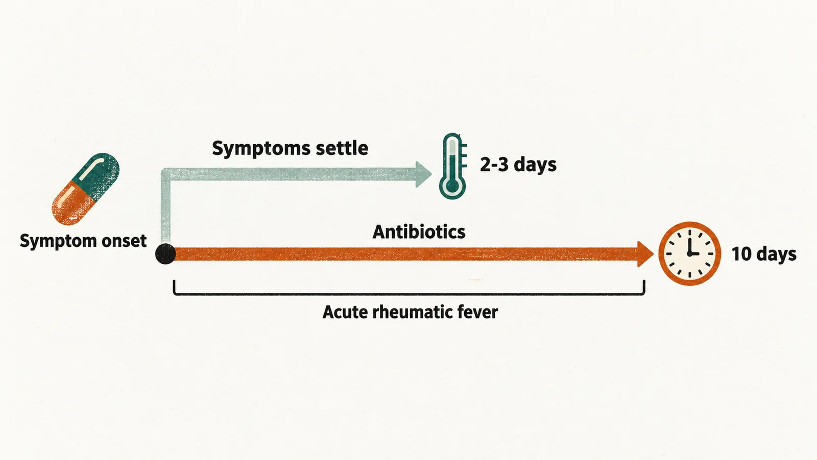
Symptoms usually settle within 2-3 days, but the course is not shortened for that reason. The full 10 days is what prevents acute rheumatic fever, and treatment started more than about 10 days after the onset of symptoms still treats the pharyngitis but no longer prevents the rheumatic complication.
For patients with a penicillin allergy, cephalexin, cefadroxil, clindamycin, azithromycin or clarithromycin are alternatives, but cephalexin and cefadroxil should be avoided in immediate-type penicillin hypersensitivity, and resistance to the macrolides and clindamycin occurs and varies geographically — so the choice is better guided by local susceptibility data than by convenience.
Public health
Treatment also bears on contagiousness: a child with scarlet fever is contagious during the acute illness, until 24-48 hours after antibiotics are started. Children should be excluded from school or childcare until they have taken appropriate antibiotics for at least 24 hours. Scarlet fever is a notifiable disease in several European countries. Post-exposure prophylaxis is not routinely recommended for household contacts.
Complications
Complications divide into those caused by spread of the infection and those caused by the immune response to it.
Suppurative complications (those that form pus) follow local or haematogenous (bloodstream) spread: cervical lymphadenitis (infected neck lymph nodes), peritonsillar and retropharyngeal abscess, otitis media, and invasive group A streptococcal disease with sepsis, cellulitis, pneumonia, septic arthritis or osteomyelitis.
Immune-mediated complications appear weeks after the acute illness. Acute rheumatic fever is the one the ten-day course prevents.
- Acute rheumatic fever — the most important, and still a leading cause of acquired cardiac valve disease where access to antibiotics is limited. It typically appears about 1-5 weeks after the preceding pharyngitis or skin infection.
- Post-streptococcal glomerulonephritis — appears about 10 days after pharyngitis and up to 3 weeks after a skin infection, and is more often associated with skin infection than with pharyngitis.
- PANDAS (paediatric autoimmune neuropsychiatric disorders associated with streptococcal infections) — the obsessive-compulsive-disorder (OCD) and tic-spectrum presentation described after streptococcal infection; the entity remains debated and the evidence base is weaker than for the other two.
The shared mechanism is molecular mimicry: streptococcal antigens share epitopes (the small molecular structures that antibodies recognise) with human cardiac, renal and neurological tissue, so antibodies raised against the bacterium cross-react with host tissue.