Fifth disease, also called erythema infectiosum, is the childhood exanthem that is usually over by the time it is diagnosed. The child is well, afebrile and no longer contagious when the rash appears, which is convenient for the child and inconvenient for everyone who was exposed during the week beforehand. It is also the classic exanthem with the sharpest clinical consequence outside childhood, because parvovirus B19 stops red cell production and the people who depend on a high rate of red cell production notice it most.
The name comes from the historical numbering of the classic exanthems, in which it is the fifth, after measles, scarlet fever, rubella and the now-discarded Duke’s disease. The synonym erythema infectiosum is more descriptive, but the numbering has survived in clinical use and in the name of the illness.
A virus with a single target cell
Parvovirus B19 is a small DNA virus in the Parvoviridae family, and the smallest DNA virus known to infect humans. Its tropism, the range of cells it can infect, is unusually narrow: it infects erythroid precursors in the bone marrow, the cells that mature into red cells, above all the proerythroblasts, an early stage of that maturation. Infection arrests erythropoiesis at that stage, so the later stages of red cell maturation do not happen. Affected proerythroblasts become enlarged and die.
The consequences depend entirely on how much the person depends on ongoing red cell production:
- Healthy children compensate through the marrow’s reserve and have no haematological problem; the visible illness is the rash.
- People with chronic haemolysis — sickle cell disease, hereditary spherocytosis, thalassaemia — rely on a continuously high reticulocyte output (reticulocytes are the young red cells the marrow releases). Arresting it produces a transient aplastic crisis with rapidly worsening anaemia.
- Fetuses have a short red cell lifespan and limited reserve, so fetal erythropoiesis is shut down effectively: severe fetal anaemia can progress to high-output cardiac failure and hydrops fetalis, an abnormal accumulation of fluid in the fetus.
A biphasic illness
The two phases are separated in time, and each has its own clinical meaning.
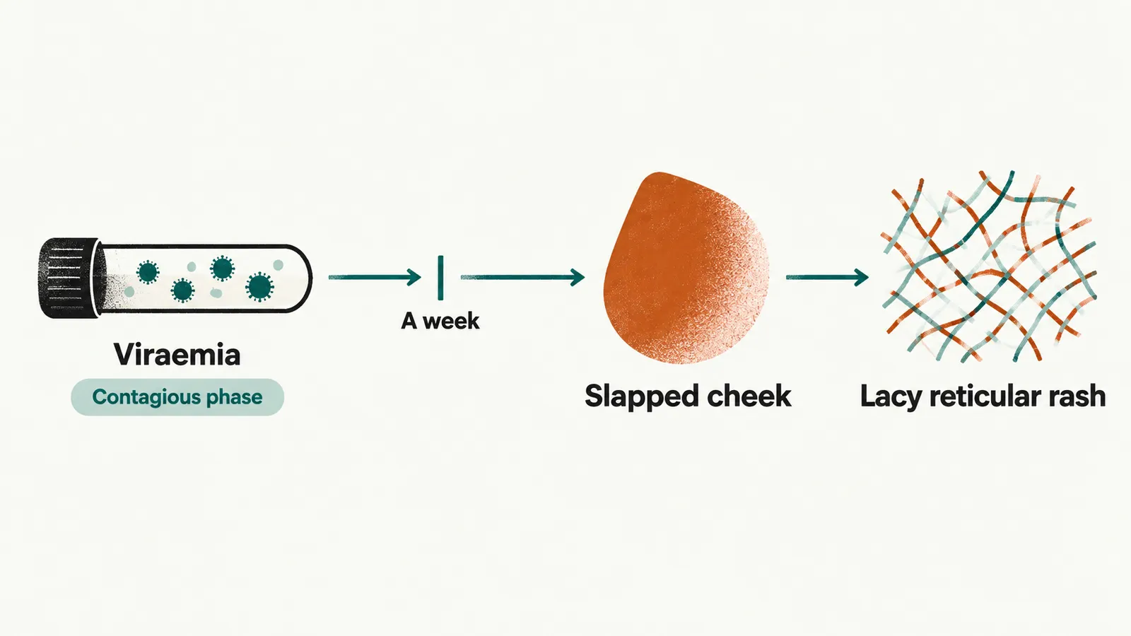
First phase: viraemia
About a week after inoculation there is an intense viraemia (virus circulating in the blood) with a non-specific flu-like illness — fever, malaise, headache, myalgia. Haematological changes appear at this stage: reticulocytopenia, a fall in haemoglobin, and sometimes leucopenia or thrombocytopenia. This is the contagious phase. Respiratory droplets spread the virus, and the child is shedding before anyone knows what the illness is.
Second phase: rash and joints
The rash follows roughly a week later and is thought to be immunologically mediated — immune-complex deposition rather than viral invasion of the skin, which is why it appears as the viraemia resolves. By this point the child is afebrile and well, and no longer infectious.
- Slapped cheek: an erythematous, well-demarcated, non-painful malar rash (over the cheekbones), often with sparing around the mouth. This is the diagnostic feature.
- Lacy reticular rash on the trunk and limbs: a macular, lacy (reticular) pattern, often more prominent on the extensor surfaces, that spares the palms and soles and is often itchy.
- Recurrence: the rash can fade and reappear over weeks, provoked by sunlight, heat, exercise or emotion. This behaviour is characteristic and is a useful diagnostic pointer.
There is no enanthem (no matching lesions on the mucous membranes) and no desquamation (peeling). In adults, particularly women, the second phase may be dominated by joints rather than skin, with a symmetric small-joint polyarthropathy of the hands, feet and knees that lasts 1-3 weeks and occasionally months. The slapped-cheek distribution can resemble the malar rash of systemic lupus erythematosus; age, the absence of scaling, and the absence of systemic features such as serositis or renal disease separate them.
Complications by population
Who develops a complication follows from the two mechanisms already described: the arrest of red cell production, which matters in proportion to how much a person depends on it, and the immune response, which produces the joint symptoms.
| Population | Complication |
|---|---|
| Healthy children | None in most cases; occasional transient arthralgia |
| Healthy adults, more often women | Polyarthropathy syndrome |
| Chronic haemolytic anaemia | Transient aplastic crisis |
| Pregnancy | Fetal anaemia and hydrops fetalis |
| Immunocompromised | Chronic anaemia, pure red cell aplasia (failure of red cell production alone) |
Transient aplastic crisis
A child with chronic haemolysis normally maintains a high reticulocyte count. Parvovirus B19 removes it. Over 5-10 days the child develops pallor, weakness and lethargy with severe anaemia, and the blood film shows the paradox that defines the condition: severe anaemia with no reticulocyte response. It is usually self-limited over 1-2 weeks, but transfusion is often required, and in most immunocompetent people it happens only once, because immunity afterwards is protective.
Infection in pregnancy
Vertical transmission occurs in approximately 17-33% of maternal infections, with the highest risk of transmission and of severe fetal effects in mid-pregnancy, and the fetus cannot mount the reticulocyte response that protects the child. Severe fetal anaemia leads to high-output cardiac failure and hydrops. When fetal anaemia or hydrops develops, intrauterine transfusion substantially improves survival — reported at 67-84% with transfusion against 30-50% without. This is the reason the diagnosis is worth making in a household with a pregnant woman, even though the child needs nothing.
The practical point follows from the biphasic structure: a pregnant woman whose child is in the febrile, flu-like phase of a parvovirus illness is being exposed at the point of maximum contagiousness, and by the time the child has slapped cheeks the window for her has usually closed. Assessment belongs to the febrile phase, not the rash.
Diagnosis
In an otherwise healthy child with a textbook slapped-cheek rash and a lacy eruption, no test is needed. Where confirmation matters — aplastic crisis, immunocompromised patients, pregnancy, fetal samples — parvovirus B19-specific IgM (recent infection) and IgG (past infection or immunity) can be measured, PCR can detect viral DNA, and immunohistochemistry can identify viral antigen in fetal or placental tissue.
Management and public health
There is no specific antiviral. Healthy children need no treatment, no isolation, and no exclusion from school, because they are no longer infectious once the rash is visible. Transfusion is used for a severe aplastic crisis, intrauterine transfusion for fetal hydrops, and intravenous immunoglobulin for chronic parvovirus B19 infection in immunocompromised patients.