
An adult who has had a urinary tract infection is rarely imaged afterwards. A child often is, because the infection may be the first visible sign of a urinary tract that was malformed before birth. Imaging after a first febrile UTI answers three separate questions, and each modality answers only one of them:
- Is there an obstruction or a structural malformation?
- Is there vesicoureteral reflux, the backflow of urine from the bladder towards the kidney?
- Is there already renal damage?
Ultrasound first
Ultrasound of the kidneys and urinary tract is the first-line study after a first febrile UTI in a young child. It is non-invasive, involves no radiation, and addresses the structural question directly. The sonographer looks for:
- Renal hypoplasia or dysplasia — a small or disorganised kidney with abnormal parenchymal thickness
- Dilatation of the renal pelvis (hydronephrosis) or of the ureter (megaureter), suggesting obstruction
- Bladder wall thickening and post-void residual urine, which suggest bladder outlet obstruction or incomplete emptying
- A duplex collecting system, an anatomical variant that often coexists with reflux
A normal ultrasound does not exclude reflux, because reflux is a dynamic event that a static image cannot show. It does substantially reduce the probability of an obstructive malformation.
Cystography — selective, not routine
Voiding cystourethrography (VCUG) is the only study that directly demonstrates reflux: contrast is instilled into the bladder through a catheter and followed on real-time X-ray (fluoroscopy) as the child voids. If contrast climbs into the ureter and renal pelvis, reflux is present and can be graded. It is also the diagnostic study for posterior urethral valves — congenital folds in the posterior urethra of boys that obstruct the outflow of urine — which appear as a dilated posterior urethra with a thickened bladder wall and dilated upper tracts in a male infant.
Because catheterisation is unpleasant and involves ionising radiation, cystography is used selectively. The decision rule that emerges from pediatric teaching rests on three findings, any one of which justifies it:
- An abnormal ultrasound — hypoplasia, abnormal echogenicity (brightness of the kidney tissue on ultrasound), pelvic or ureteral dilatation, or a bladder abnormality.
- A non-E. coli organism — Klebsiella, Pseudomonas or Proteus, which raises the probability of a structural abnormality.
- Recurrent febrile UTI — a second or third febrile infection, even after a normal ultrasound and a first E. coli infection.
The combination matters more than any single item. A normal ultrasound with an E. coli first infection does not by itself call for a cystogram; the same normal ultrasound with a Pseudomonas infection does. Current European urology guidance is candid that the criteria for selecting children for reflux detection are supported only weakly, and that renal ultrasound plus clinical risk factors (age, sepsis, white cell count, abnormal ultrasound) can be combined into predictive scores rather than used as rigid thresholds.
DMSA — the scar scan
Dimercaptosuccinic acid (DMSA) scintigraphy, a nuclear scan using a radioactive tracer, answers the third question. The tracer is taken up by functioning tubular cells, so areas of scarring appear as focal photopenic defects — patches where no tracer has been taken up. The decisive scan is done 4–6 months after the infection, once acute inflammation has resolved and any defect can be called permanent; a scan taken during the acute illness shows parenchymal involvement but cannot distinguish a lesion that will heal from one that will scar. DMSA localises damage but does not explain it — that is the role of cystography.
MAG-3 — drainage and split function
Mercaptoacetyltriglycine (MAG-3) scintigraphy is a dynamic study used when the question is drainage rather than cortical scarring: suspected ureteropelvic junction obstruction (a blockage where the renal pelvis meets the ureter), or a dilated system where it matters whether urine empties. It reports the differential function of each kidney as a percentage — the share of total kidney function that each side contributes, also called split function. DMSA and MAG-3 can both give split function; the practical difference is drainage versus focal cortical defects.
Findings worth recognising
Across these studies, a small set of findings recurs, and each points towards a different underlying problem:
| Finding | Study | What it means |
|---|---|---|
| Focal photopenic defect | DMSA, 4–6 months after infection | Permanent scarring; about 15% of children after a first UTI, higher after recurrent febrile episodes |
| Reflux, graded I–V | Cystography | Retrograde urine flow from bladder to kidney; idiopathic or secondary |
| Posterior urethral valves | Cystography in a male infant | Urethral obstruction with high-pressure secondary reflux — one of the most damaging structural abnormalities |
| Hydronephrosis | Ultrasound | Obstruction, reflux, megaureter, or transient dilatation |
| Duplex collecting system | Ultrasound | Two collecting systems, each draining one part (moiety) of the kidney; the upper moiety classically obstructs and the lower moiety classically refluxes |
| Megaureter | Ultrasound | A dilated ureter, from obstruction, reflux or both |
The ladder in practice
Putting the studies in order gives a ladder that escalates only when a finding or the clinical course justifies it:
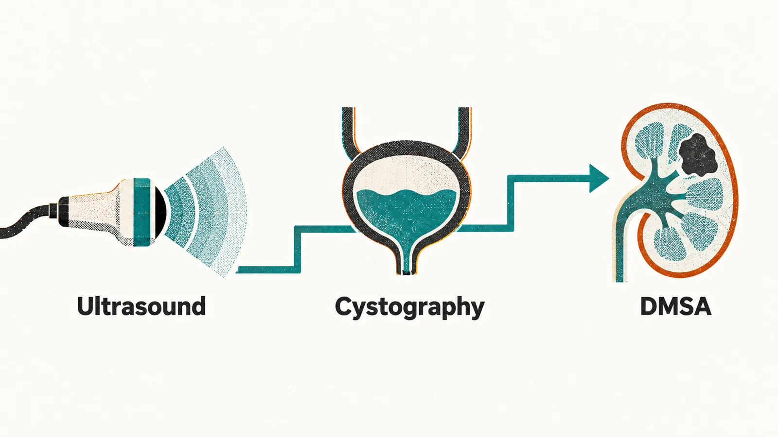
- First febrile UTI in a young child → renal and urinary tract ultrasound.
- Abnormal ultrasound → cystography, plus a delayed DMSA to establish a baseline for scarring.
- Normal ultrasound but a non-E. coli organism or a recurrent febrile UTI → cystography, plus delayed DMSA.
- Normal ultrasound, single E. coli infection, no recurrence → delayed DMSA is the study that matters, and cystography is usually deferred.
Where that ladder is applied differs between guidelines. A bottom-up strategy (ultrasound, then cystography if indicated, then DMSA) starts with the least invasive study and escalates. A top-down strategy starts with acute DMSA to confirm parenchymal involvement and proceeds to cystography only if the kidney is affected; this has become more common in European practice because it exposes fewer children to catheterisation and radiation. Both strategies are in use, and the choice belongs to the local protocol.
Recurrence appears on the ladder as a reason for cystography. Yet in a toilet-trained child whose infections keep returning, the explanation often lies less in the anatomy than in how the bladder and bowel behave between infections.
At a glance
- Ultrasound first, for structure. It cannot exclude reflux.
- Cystography is selective: abnormal ultrasound, non-E. coli organism, or recurrent febrile UTI.
- Cystography is how posterior urethral valves are diagnosed.
- DMSA at 4–6 months shows permanent scarring; acute DMSA shows involvement that may or may not resolve.
- MAG-3 answers drainage and split function.
- Bottom-up and top-down strategies are both current practice; the local protocol decides which is used.