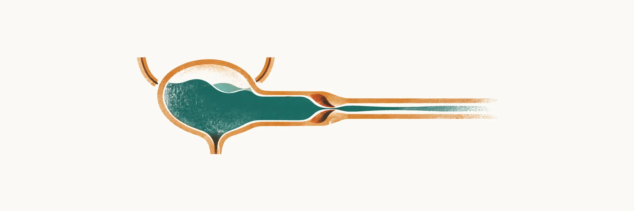
An obstruction anywhere along the urinary tract raises the pressure behind it, and in a fetus or infant that pressure falls on a kidney that is still being built. Obstructive uropathy is the resulting pattern of dilatation and parenchymal damage. Within the group of malformations known as congenital anomalies of the kidney and urinary tract (CAKUT), the most damaging example is a pair of mucosal folds in the posterior urethra of boys, and its consequences extend from before birth into adult life.
What obstruction does to a kidney
Two mechanisms run in parallel, and both are reasons to relieve an obstruction rather than observe it.
Pressure. Urine that cannot leave the system dilates the tract behind the blockage — the renal pelvis first, then the ureters, then the bladder — and sustained high pressure damages the developing parenchyma. Sterile urine alone can do this, which is why dysplasia, abnormally formed kidney tissue, is sometimes found in kidneys draining a high-pressure system before any infection has occurred.
Infection. Stasis leaves urine in place to be colonised, so an obstructed tract infects more easily. It also makes the infection more consequential: a febrile urinary tract infection (UTI) in a child with an obstruction is a signal that the obstruction itself needs attention, not only an antibiotic.
Obstruction in children occurs at a few characteristic levels: the ureteropelvic junction, where the renal pelvis meets the ureter; the ureterovesical junction, which is also the site of the valve mechanism that fails in vesicoureteral reflux, the backflow of urine from the bladder towards the kidney; and the posterior urethra, where valves in boys produce the most severe form.
Posterior urethral valves
Posterior urethral valves (PUV) are congenital mucosal folds in the posterior urethra of a boy that obstruct the outflow of urine. They are one of the few life-threatening congenital anomalies of the urinary tract found in the neonatal period. PUV account for roughly 60% of congenital lower urinary tract obstruction, their incidence is estimated at 1 in 7,000–8,000 live births, and up to 17% of paediatric end-stage renal disease — kidney failure requiring dialysis or transplantation — is attributed to them.
The commonest form, Young type I, is a ridge continuous with the verumontanum (a small mound on the floor of the posterior urethra) that divides into two fork-like processes, and it accounts for 90–95% of cases. Because the obstruction sits below the bladder, everything above it changes: nearly all boys with PUV have dilated upper tracts, secondary vesicoureteral reflux is present in at least 50%, and the bladder wall and bladder neck hypertrophy. A kidney draining a high-grade refluxing system in this setting often functions poorly.
Before and after birth
Antenatal ultrasound raises the suspicion in most cases. The findings are a distended, thick-walled bladder with bilateral hydroureteronephrosis (dilatation of both ureters and both renal collecting systems), sometimes with the “keyhole” appearance of the dilated posterior urethra. Severe obstruction reduces fetal urine output and produces oligohydramnios, too little amniotic fluid, which matters because amniotic fluid is needed for lung development.
None of these signs is decisive on its own. A thick-walled bladder predicts PUV better than the keyhole sign, and the imaging is not specific — obstructive and non-obstructive dilatation can look alike — so a suspected lower urinary tract obstruction is normally referred to a centre with both prenatal and postnatal expertise. Restoring amniotic fluid with a prenatal shunt, a drain from the fetal bladder into the amniotic cavity, may improve early survival, but the randomised PLUTO trial found no long-term benefit for renal function.
After birth the picture depends on how severe the obstruction is. A boy may have a poor urinary stream, a palpable bladder, failure to thrive, or a urinary tract infection that behaves like sepsis; blood tests may show impaired kidney function with electrolyte disturbance. Undescended testis and inguinal hernia are more common in these boys than in others.
Confirming the diagnosis
Creatinine, urea and electrolytes are followed closely through the first days, and the initial management involves a paediatric nephrologist as well as a urologist. Voiding cystourethrography (VCUG), with lateral views of the urethra during voiding and no catheter in place, is the study that defines the anatomy: a dilated posterior urethra, a thick-walled bladder and dilated upper tracts. Nuclear renography with dimercaptosuccinic acid (DMSA) or mercaptoacetyltriglycine (MAG-3) then assesses how much function each kidney contributes and whether either has already scarred.
Relieving the obstruction
Drainage comes first and definitive treatment second, and the order matters because the infant’s chemistry and kidneys must be stabilised before instrumentation.
- Drain the bladder with a transurethral or suprapubic catheter and start antibiotic prophylaxis.
- Confirm the anatomy on VCUG during the voiding phase of the study, with the bladder already drained.
- Ablate the valves endoscopically once the infant is stable, provided the urethra admits the instruments. Extensive electrocoagulation is avoided, because urethral stricture is the commonest complication of the procedure.
- Maintain drainage when the child is too small for endoscopy, or when ablation fails to decompress the upper tracts or improve renal function. A vesicostomy, an opening of the bladder onto the lower abdominal wall, is one option and stabilises or improves the upper tracts in up to 90% of cases; high urinary diversion, which drains urine from above the bladder, is reserved for children whose upper tracts do not decompress with bladder drainage alone.
Diversion is not a more renoprotective treatment than ablation. A systematic review of comparative studies found that the worse kidney outcomes reported after diversion were largely explained by worse kidney function at presentation, so the choice between the two is usually anatomical and practical rather than a matter of protecting the kidneys.
The valve bladder and lifelong follow-up
Relieving the obstruction does not end the problem, because a bladder that developed under high pressure continues to misbehave. Valve bladder describes that dysfunction — poor compliance (the bladder cannot fill without its pressure rising) or small capacity, detrusor overactivity (involuntary contractions of the bladder wall muscle during filling), and later myogenic failure, in which that muscle no longer empties the bladder effectively — and it can progress long after anatomically successful ablation. Daytime and night-time continence are often delayed. Management is guided by urodynamic assessment, which measures bladder pressure and flow during filling and voiding, where indicated. It may include anticholinergic therapy for overactivity, alpha-blockers or clean intermittent catheterisation (emptying the bladder with a catheter at set times) when emptying is poor, and nocturnal drainage when a high overnight urine output distends the bladder.
Renal prognosis is set largely by the damage present at diagnosis, and the most useful single marker is the nadir creatinine — the lowest serum creatinine during the first year after decompression. A nadir creatinine above 0.85 mg/dL (75 µmol/L) is correlated with a poor renal prognosis, and published cut-offs range from 0.85 to 1.2 mg/dL. Estimates of eventual kidney failure vary with the cohort studied: a systematic review reported chronic kidney disease in about 32% and end-stage renal disease in about 20%, while the European guideline notes chronic kidney disease in up to 65% of patients in some series. Because bladder function can deteriorate years after ablation, both bladder and kidney function are monitored lifelong.
At a glance
- Obstruction damages the kidney by pressure and by infection, and sterile high pressure alone can leave dysplasia.
- PUV are congenital folds in the posterior urethra of boys, with an incidence of 1 in 7,000–8,000 live births, and account for roughly 60% of congenital lower urinary tract obstruction.
- Antenatal suspicion comes from a thick-walled bladder with bilateral hydroureteronephrosis and oligohydramnios; the diagnosis is confirmed after birth on voiding cystourethrography.
- Treat with bladder drainage and antibiotic prophylaxis first, then endoscopic valve ablation; extensive coagulation risks a urethral stricture.
- A nadir creatinine above 0.85 mg/dL (75 µmol/L) is associated with a poor renal prognosis, and in one systematic review about one boy in five reached end-stage renal disease.
- Valve bladder dysfunction can progress after successful ablation, so bladder and kidney function are followed for life.