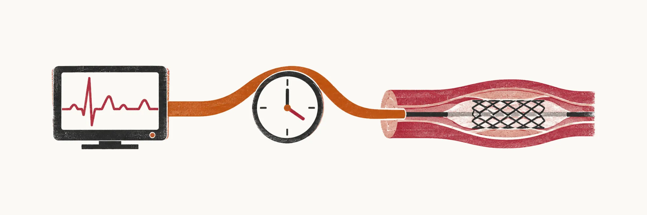
The first hour after presentation decides the trajectory of an acute myocardial infarction, and the aim of that hour is not simply to make a diagnosis. It is to identify the patient whose artery is closed, move that patient to reperfusion — reopening the artery, by primary percutaneous coronary intervention (PCI) with balloon and stent or by fibrinolysis, a clot-dissolving drug — and recognise the patient who is about to decompensate for a different reason: shock, pulmonary oedema, a life-threatening rhythm.
The first ten minutes
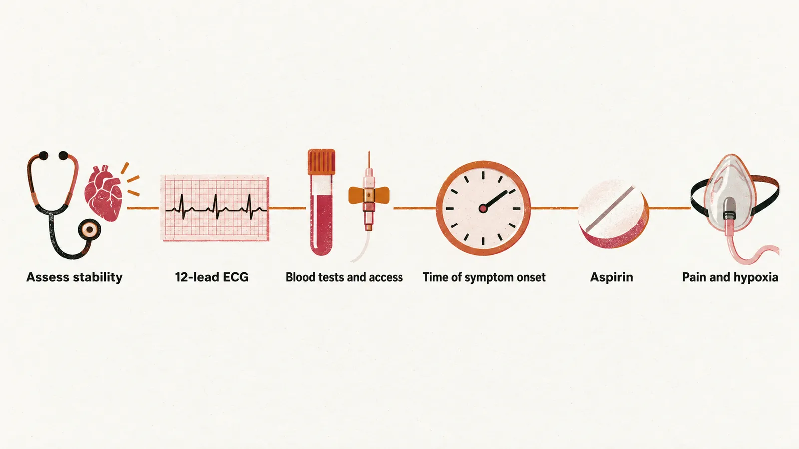
The first ten minutes cover six tasks, from checking stability to the treatment that can start before any result returns.
- Assess stability. Airway, breathing, circulation. A patient in shock, pulmonary oedema or cardiac arrest needs the catheterisation laboratory and intensive support regardless of ECG subtleties.
- Obtain and read a 12-lead ECG within 10 minutes of first medical contact. Look for ST elevation in two contiguous leads (leads that view the same wall), the aVR pattern (ST elevation in lead aVR with widespread ST depression, which suggests left main or severe three-vessel disease), and reciprocal changes (ST depression that mirrors an elevation in the opposite wall). ST elevation defines ST-elevation myocardial infarction (STEMI), the pathway to immediate reperfusion. A new left bundle branch block masks the ST segment and is no longer a trigger by itself; that patient is managed through the non-ST-elevation pathway unless the instability or ongoing ischaemic symptoms demand otherwise.
- Secure intravenous access and send blood for troponin, full blood count, creatinine and coagulation, without waiting for results before treating.
- Take the history that changes management. Time of symptom onset determines whether reperfusion is still worthwhile and which strategy is available; bleeding history, anticoagulant use, recent surgery, stroke and malignancy determine whether fibrinolysis is safe.
- Give aspirin to every patient without a contraindication. It is the one drug that does not depend on the ECG result.
- Treat pain and hypoxia while the pathway runs: nitrates for ongoing angina, morphine for severe pain, oxygen only if saturation falls below 90%.
Killip classification
Killip class is a bedside estimate of how much haemodynamic compromise the infarct has caused, and it predicts short-term mortality better than infarct size estimates from the ECG.
| Class | Bedside findings | 30-day mortality (STEMI treated with PCI) |
|---|---|---|
| I | No clinical signs of heart failure | 3% |
| II | Crackles in the lung fields, third heart sound, raised jugular venous pressure | 14.6% (classes II–III combined) |
| III | Frank pulmonary oedema | 14.6% (classes II–III combined) |
| IV | Cardiogenic shock — hypotension with hypoperfusion | 47.5% |
The pattern across the classes is the point. Reperfusion, modern intensive care and mechanical support have brought mortality in the milder classes down to single figures, while cardiogenic shock remains close to a coin flip. Those figures come from a national registry of STEMI patients treated with PCI, so they describe that population rather than ACS in general, and definitions of shock vary between studies — which is why the numbers are best read as magnitudes.
Killip class is not the same as the New York Heart Association (NYHA) functional classification. Killip describes acute haemodynamic state during infarction and predicts early mortality; NYHA describes chronic functional limitation in established heart failure. Class should be assigned at first assessment and reassessed after reperfusion, because a patient who arrives in class III and improves to class II after PCI is a different prognostic case from one who stays in class III.
From emergency department to catheterisation laboratory
When the ECG shows definite ST elevation in two contiguous leads, the reperfusion pathway activates: notify the interventional team, give the P2Y12 inhibitor (the second antiplatelet drug, added to aspirin) and parenteral anticoagulant as described in Antithrombotic and Symptom Pharmacotherapy in ACS, and transfer with a target of first-medical-contact-to-device time of 90 minutes when PCI is on site or 120 minutes when transfer is needed. If the hospital has no PCI capability and the target cannot be met, the choice between immediate fibrinolysis and transfer for PCI follows the rules and time windows set out in Reperfusion in Acute Myocardial Infarction.
While the patient waits, three things run in parallel: continuous rhythm monitoring, because the first hours carry the highest risk of ventricular arrhythmia; symptom and haemodynamic control; and preparation for the possibility of deterioration, with the defibrillator immediately available.
In the catheterisation laboratory, the culprit lesion — the one responsible for the infarction — is identified and stented, with radial access through the wrist preferred. When multivessel disease is found in a stable patient, complete revascularisation — during the index procedure or within 45 days — is the current recommendation; in cardiogenic shock, only the infarct-related artery is treated at once.
For patients without ST elevation, the pathway is different in kind. Aspirin and parenteral anticoagulation start immediately, the P2Y12 inhibitor is generally held until the coronary anatomy is known, and the timing of angiography is set by risk category rather than by a fixed protocol.
Cardiogenic shock
Cardiogenic shock is hypotension with hypoperfusion caused by the failing heart — Killip class IV. Shock complicates about 10% of ST-elevation infarctions and carries early mortality of 40% to 50%, so the pathway bends around it. The patient needs the catheterisation laboratory and circulatory support at the same time, not one after the other.
- Revascularisation. Only the infarct-related artery is treated at the index procedure, because additional lesions add procedure time and risk without proven benefit in that setting; the 2023 European guideline gives infarct-related-artery-only PCI a class I, level B recommendation. Early revascularisation remains the intervention with the strongest evidence.
- Mechanical support. The 2025 American guideline treats a microaxial flow pump, a small catheter-mounted pump that supports the output of the left ventricle, as reasonable in selected patients, restricted to those matching the population in which it was tested: ST-elevation infarction with shock within 24 hours, not comatose, SCAI stage C to E (the more severe stages of the Society for Cardiovascular Angiography and Interventions shock classification), and adequate peripheral vasculature. In that group, death at 180 days fell from 58.5% to 45.8% — a number needed to treat of 8 — at the cost of more bleeding, limb ischaemia and renal replacement therapy. The 2023 European guideline predates that trial and is more reserved about mechanical support, so its position should be read as the older one.
- Supportive care. Continuous rhythm monitoring, blood pressure and urine output, and preparation for renal replacement or ventilation as the clinical course dictates.
The failing ventricle itself — why shock kills, and how it fits the rest of the post-infarction picture — is described in Complications of Myocardial Infarction.
The first 24 hours after reperfusion
Post-procedure care is mostly about detecting what the procedure did not fix. The heart is watched first, then the consequences of the procedure itself, and the drugs are adjusted to the new situation.
Rhythm needs interpretation rather than reflex. Reperfusion arrhythmias are common, typically slow, wide-complex and self-limiting, and usually need no treatment; they are a sign that flow has been restored. Sustained ventricular tachycardia or fibrillation is a different matter and requires treatment. Continuous monitoring therefore continues after the procedure.
Haemodynamics — blood pressure, heart rate and urine output — are followed for heart failure or shock. Serial examination and, where indicated, echocardiography look for mechanical complications.
The procedure leaves its own marks. The access site is checked for bleeding, haematoma and pseudoaneurysm (a leak from the punctured artery contained by surrounding tissue), which are more frequent after femoral access. Troponin shows a rise-and-fall pattern that confirms the diagnosis, and the peak gives a rough indication of infarct size. Creatinine, potassium and haemoglobin are followed because contrast, diuretics and bleeding all interact with them.
Antithrombotic therapy changes once the artery is stented: dual antiplatelet therapy (DAPT, aspirin plus a P2Y12 inhibitor) continues; routine post-procedure anticoagulation does not, unless a separate indication exists.
Starting secondary prevention before discharge
The discharge prescription is where long-term survival is decided, and it starts in hospital rather than at the first outpatient review. The minimum for the acute admission is a high-intensity statin for every patient regardless of baseline low-density lipoprotein (LDL) cholesterol, with beta-blocker, renin–angiotensin system blockade (an ACE inhibitor or related drug) and mineralocorticoid receptor antagonism (spironolactone or eplerenone) added according to ejection fraction, infarct location and comorbidity. The indications, doses and laboratory caveats are covered in Post-MI Long-Term Management; the organising principle is that the weeks after infarction are when the ventricle remodels and the risk of a second event is highest.