
Reperfusion means restoring flow through an occluded coronary artery. In ST-elevation myocardial infarction (STEMI), where the ECG generally reflects an acutely occluded artery, it is the only intervention that changes the fate of infarcting myocardium, and the decision it rests on is which strategy can open the artery soonest. Time targets, drug selection, post-treatment angiography and late presentation all follow from that question.
There are two strategies. Primary percutaneous coronary intervention (PCI) opens the artery mechanically in the catheterisation laboratory as the first reperfusion treatment; fibrinolysis gives a drug that dissolves the clot. Primary PCI is the default when it can be delivered promptly, because it achieves better and more durable flow than fibrinolysis and avoids systemic fibrinolysis with its bleeding and stroke risk. Fibrinolysis remains the alternative when the geography or the clock makes timely PCI impossible, and it works best when it is started early.
Time targets
| Target | Interval |
|---|---|
| First medical contact (FMC) to 12-lead ECG | ≤10 minutes |
| FMC to primary PCI (device) where PCI is on site | ≤90 minutes |
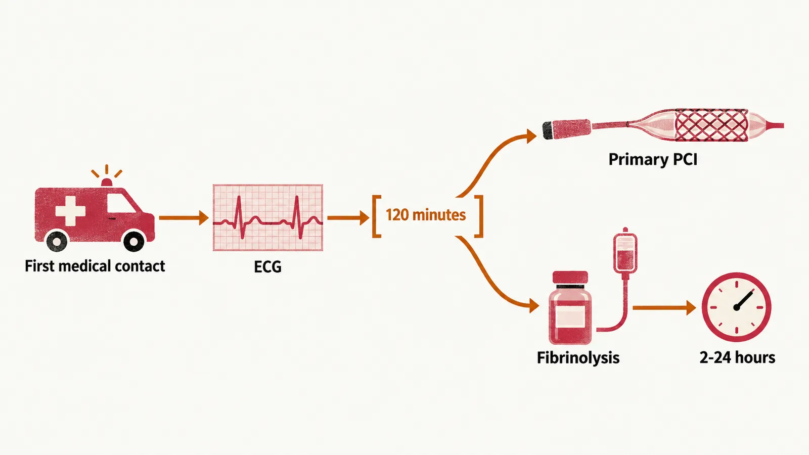
| FMC to primary PCI where transfer is required | ≤120 minutes |
| Onset of fibrinolysis, when PCI cannot be delivered in time | as soon as possible, ideally within 10 minutes of the STEMI diagnosis |
| Routine angiography after successful fibrinolysis | 2–24 hours |
The 120-minute figure is not an alternative target but the decision point. If primary PCI can be delivered within that 120-minute window, transfer for PCI is the strategy. If it cannot, fibrinolysis is started first, and transfer follows — fibrinolysis should not be delayed by waiting for transfer to a PCI centre.
Primary PCI
In primary PCI, a catheter is passed from an artery in the wrist or groin to the opening of the coronary artery, a guidewire crosses the occlusion, the lesion is prepared with balloon angioplasty, and a stent is deployed.
Access is radial (through the wrist) whenever the anatomy allows. Compared with the femoral route through the groin it reduces access-site bleeding, retroperitoneal haematoma and vascular complications, and it is the approach recommended by current European guidance. Drug-eluting stents are standard: they reduce restenosis, the renarrowing of the treated segment, compared with bare metal stents, at the cost of delayed endothelialisation — the slower growth of the vessel lining over the stent. That means the stent surface stays thrombogenic for longer and antiplatelet cover has to last until it is covered.
Routine thrombus aspiration, sucking clot out through a catheter before stenting, is not recommended. It may be used selectively when a large thrombus burden threatens distal embolisation during stent deployment — in practice, in about 4% to 7% of primary PCI procedures — but the randomised evidence does not support routine use.
When angiography shows multivessel disease, the strategy depends on stability. The culprit lesion is the one that caused the infarction, in the infarct-related artery; the others are non-culprit lesions. In a haemodynamically stable STEMI patient, complete revascularisation — treating significant non-culprit lesions as well, either during the index procedure or within 45 days — carries a Class I recommendation in the 2023 European guideline, on the strength of trials showing fewer subsequent ischaemic events than with culprit-only treatment. In cardiogenic shock the logic reverses: only the infarct-related artery is treated immediately, because additional lesions add procedure time and risk without proven benefit in that setting.
When the artery is open but the muscle is not reperfused
An open epicardial artery does not guarantee that blood reaches the muscle it supplies. No-reflow — slow or absent flow after stenting despite a patent vessel — is caused by microvascular obstruction: clot and debris embolised downstream during the procedure, injury caused by reperfusion itself, and differences in how individual microcirculations respond. Angiographic no-reflow is seen in more than 20% of primary angioplasty cases, although reported rates vary widely with the definition used, and it is associated with a larger infarct, worse left ventricular function and higher mortality.
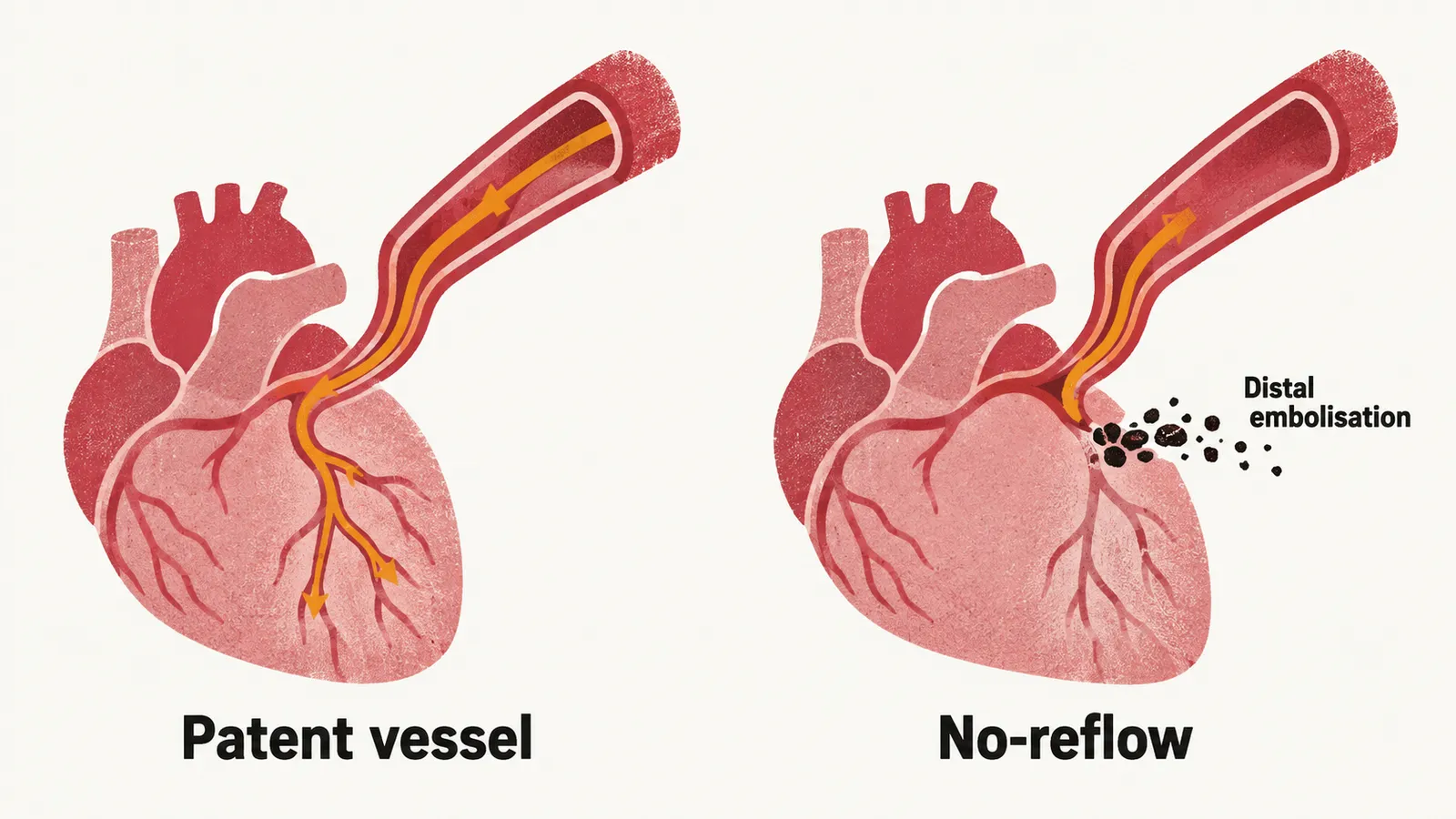
That is why an angiographic result is not the same as a good outcome, and why distal embolisation is treated as a catheterisation-laboratory problem rather than an incidental finding. Glycoprotein IIb/IIIa inhibitors are the bailout drugs when flow is poor or a thrombotic complication appears during the procedure, and their use is otherwise not routine (see Antithrombotic and Symptom Pharmacotherapy in ACS). The mechanisms behind microvascular obstruction are described in Pathophysiology of Myocardial Infarction.
Fibrinolysis
When primary PCI cannot be delivered in time, the clot is attacked with drugs instead. Fibrinolysis activates plasminogen to plasmin, which digests the fibrin scaffold of the thrombus. Its benefit is steeply time-dependent: the largest absolute reductions in mortality occur when it is given in the first two to three hours after symptom onset, and the benefit falls as the delay lengthens. The collaborative overview of the large randomised trials found that the excess of strokes attributable to treatment is small compared with the mortality benefit, but it is real and is concentrated in the first day.
Agent choice. Fibrin-specific agents, which act mainly on plasminogen bound to fibrin in the clot, are preferred over streptokinase for better efficacy and fewer adverse reactions:
- Tenecteplase — a single weight-adjusted intravenous bolus, equivalent efficacy to alteplase with fewer non-cerebral bleeds, and the easiest to give.
- Alteplase (tPA) — fibrin-specific, given as an accelerated infusion.
- Streptokinase — non-fibrin-specific and antigenic, so repeat exposure risks allergic reactions and antibodies can blunt a second dose.
Contraindications follow the bleeding risk of systemic lysis.
- Absolute: previous intracranial haemorrhage, ischaemic stroke within the past 6 months, known cerebral arteriovenous malformation or aneurysm, major surgery or significant trauma within 3 weeks, gastrointestinal bleeding within the past month, and known bleeding disorder.
- Relative: ongoing therapeutic anticoagulation, uncontrolled severe hypertension, and active infective endocarditis.
Intracranial haemorrhage complicates roughly 1% of fibrinolysis, and the risk is higher with age 75 years or older, female sex, low body weight, previous cerebrovascular disease, and high admission blood pressure.
Angiography after fibrinolysis
Fibrinolysis is not the end of the pathway, because angiography is needed regardless of how the patient looks. What changes is how soon.
- Immediate angiography if reperfusion appears to have failed (less than 50% resolution of ST elevation at 60–90 minutes), if heart failure or shock develops, if there is electrical or haemodynamic instability, or if ischaemia recurs.
- Otherwise angiography between 2 and 24 hours after successful fibrinolysis.
Presentation after the 12-hour mark
Both strategies are built around the time since symptom onset, because muscle is still being lost while the artery stays closed. Patients presenting more than 12 hours after symptom onset need individual assessment rather than reflex intervention:
- Between 12 and 48 hours, PCI can be considered when there is evidence of ongoing ischaemia — recurrent pain, dynamic ECG changes, instability or heart failure.
- Beyond 48 hours in a stable, asymptomatic patient, routine PCI of a persistently occluded infarct-related artery is not indicated. The territory is already infarcted, and reopening it has not improved outcomes in randomised trials. These patients are managed like chronic total occlusion: revascularisation is considered when there is documented viable myocardium or symptoms attributable to that territory.
Timing of angiography in non-ST-elevation ACS
Reperfusion as an emergency belongs to ST elevation, where the artery is presumed closed. Without ST elevation there is no automatic catheterisation trigger, so the question becomes when to perform angiography, and urgency is graded by risk. The GRACE score used in the table forecasts death and reinfarction:
| Risk | Features | Timing |
|---|---|---|
| Very high | Haemodynamic instability or cardiogenic shock, recurrent or refractory chest pain despite treatment, life-threatening arrhythmia, mechanical complication, ST elevation in aVR with widespread ST depression | Immediate, within 2 hours |
| High | Rising troponin, dynamic ST–T changes, GRACE score above 140 | Within 24 hours |
| Intermediate | Diabetes, renal impairment, reduced ejection fraction, prior revascularisation, GRACE score above 109 without high-risk features | During the index hospitalisation |
| Low | No recurrent symptoms, no high-risk features, negative serial troponins | Selective, ischaemia-guided |
Two points about the high-risk row. The 24-hour timing is a considered recommendation rather than a strong one: the 2023 European guideline revised the early invasive strategy for these high-risk patients, including those with a GRACE score above 140, to class IIa, having previously graded it higher. And the aVR pattern is easy to read past: ST elevation in aVR together with ST depression in six or more other leads points toward left main or severe three-vessel disease and places the patient in the immediate group even when the rhythm and blood pressure are stable.