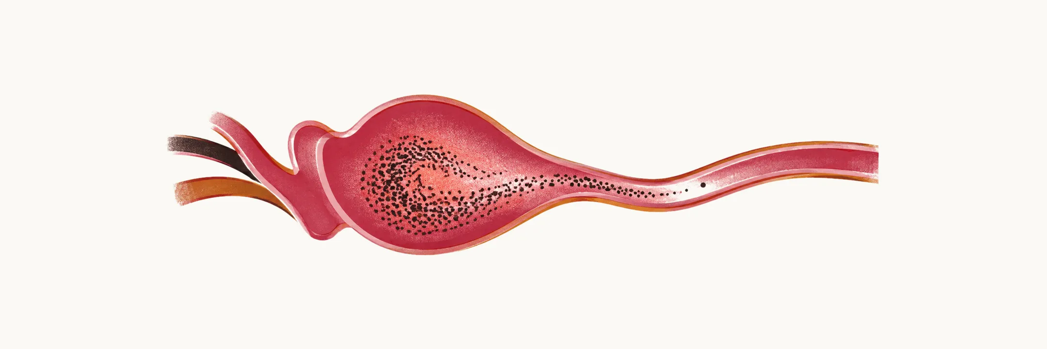
The consequences of an occluded coronary artery spread well beyond the infarcted segment. What makes them manageable is timing: arrhythmias and pump failure dominate the first hours to days, mechanical rupture clusters in the first week, and aneurysm, embolism and the later form of pericarditis belong to the weeks and months that follow. Reading a deteriorating patient with that timeline in mind narrows the differential quickly.
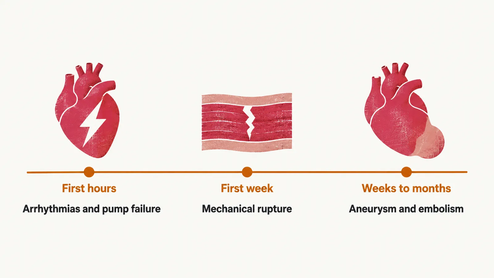
The first hours: rhythm and pump
In the first hours, two things go wrong: the injured muscle becomes electrically unstable, and the muscle that has stopped contracting can no longer maintain output.
Rhythm
Ventricular tachycardia and fibrillation. The ischaemic border zone around an infarct, the partly perfused muscle at its edge, is an electrically unstable substrate, and re-entry through it — an impulse circling through slowly conducting tissue and re-exciting the ventricle — can produce sustained ventricular tachycardia or fibrillation. In contemporary series of ST-elevation myocardial infarction (STEMI) treated with primary percutaneous coronary intervention (PCI), in which the artery is reopened with balloon and stent, these arrhythmias occur in roughly 3–7% of patients, with almost all events inside the first 24 to 48 hours. The strongest treatment is urgent reperfusion, because the rhythm is driven by ongoing ischaemia; beta-blockade reduces the risk where it is not contraindicated; defibrillation restores the rhythm, and intravenous amiodarone is the antiarrhythmic of choice when electrical therapy is insufficient. Their occurrence increases in-hospital mortality substantially, but patients who survive to discharge after an early arrhythmia have a prognosis similar to those who never had one — which is why an arrhythmia in the first 48 hours is not by itself an indication for a defibrillator.
Non-sustained ventricular tachycardia — runs lasting under 30 seconds that terminate spontaneously — is common in the first hours to days. It calls for continued monitoring and attention to ischaemia rather than routine antiarrhythmic therapy.
Reperfusion arrhythmias are a different phenomenon and should not be treated as a complication. When flow is restored they appear as slow, wide-complex, self-limiting rhythms, typically an accelerated idioventricular rhythm, a regular rhythm arising in the ventricles, and they are a sign that the vessel has reopened. They need treatment only if they cause haemodynamic compromise.
Bradyarrhythmias and atrioventricular block. In inferior infarction the right coronary artery often supplies the atrioventricular node, so bradycardia and transient high-grade block are common, usually responsive to atropine and usually transient as the artery reopens. Complete heart block in anterior infarction means something else entirely: extensive septal necrosis with loss of the conduction tissue below the node, which carries a poor prognosis and often requires pacing.
Pump
Acute heart failure and cardiogenic shock. Loss of contractile muscle raises filling pressures and lowers output, producing pulmonary congestion at one end of the spectrum and shock at the other. Cardiogenic shock complicates roughly 5–10% of infarctions and remains the leading cause of in-hospital death, with 30-day mortality close to 50% in contemporary registries — a figure that has barely moved despite reperfusion and mechanical support. This is where Killip classification, the bedside grading of heart failure during infarction, does its prognostic work, and where early revascularisation of the infarct-related artery has the strongest evidence; the decision sequence for the shocked patient, including mechanical support, is set out in the same note.
Right ventricular infarction
One form of early pump failure needs the opposite treatment. Right ventricular infarction means the right coronary artery has occluded before its right ventricular branches, so it accompanies an inferior infarct rather than an anterior or lateral one. The right ventricle is a thin-walled, low-pressure chamber that depends on venous return, and when it fails the patient becomes preload-dependent, reliant on the filling pressure delivered by venous return: systemic blood pressure falls, jugular venous pressure rises, and the lungs stay clear, because the failing chamber cannot fill the pulmonary circulation. Right precordial leads (V3R and V4R) are recorded in a suspected inferior infarct to detect it, and posterior extension is looked for with V7 to V9.
The trap is treatment. Nitrates and diuretics lower preload, and in this patient that empties the ventricle doing the work. Both current guidelines advise against nitrates when right ventricular infarction is suspected — the 2025 American guideline lists suspected right ventricular infarction, a systolic pressure below 90 mmHg, or a fall of more than 30 mmHg below baseline as reasons to avoid them — and the patient with inferior infarction and hypotension is supported with volume rather than preload reduction. Volume loading for right ventricular hypotension is conventional bedside practice rather than a guideline-level recommendation, and the nitrate prohibition itself has been challenged by a review arguing that the underlying studies were small and mixed inferior infarcts with inferior-plus-right-ventricular infarcts.
Days one to seven: mechanical rupture
Over the first week a further danger appears in the structure of the wall itself, while dead muscle has not yet been replaced by scar. Mechanical complications are rarer than they used to be and no less lethal when they occur. Population data covering 3.95 million STEMI and 5.11 million non-ST-elevation (NSTEMI) admissions found mechanical complications in 0.27% of STEMI and 0.06% of NSTEMI hospitalisations, with in-hospital mortality of 42.4% after STEMI and 18.0% after NSTEMI and no improvement across the study period. In other words, reperfusion has made these events less frequent without making them less dangerous, and they remain concentrated in patients whose artery was reperfused late or not at all.
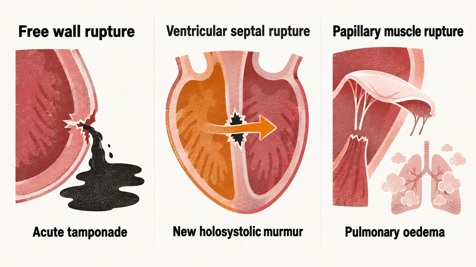
Three lesions account for most of them, and each announces itself differently:
- Free wall rupture. The necrotic ventricular wall tears, blood enters the pericardial space, and the patient develops acute tamponade, compression of the heart by blood in the pericardium, classically collapsing with pulseless electrical activity. Survival depends on immediate recognition and surgery.
- Ventricular septal rupture. A hole forms between the ventricles, producing acute right ventricular volume overload, a new harsh holosystolic murmur and cardiogenic shock. Echocardiography confirms it; surgical or percutaneous closure is required.
- Papillary muscle rupture. Loss of the muscle anchoring the mitral valve causes acute severe mitral regurgitation with sudden pulmonary oedema and hypotension. The posteromedial papillary muscle is affected more often than the anterolateral, because it has a single blood supply from the posterior descending artery, while the anterolateral muscle is usually supplied by both the left anterior descending and circumflex arteries.
Pericarditis, Dressler syndrome or reinfarction
Chest pain that returns after an infarction raises a different question. Three problems present with chest pain, a changing ECG and a raised troponin after an infarction, and timing is what separates them.
- Early pericarditis appears 1 to 3 days after a transmural infarct. It is presumed to be inflammatory, caused by inflammation in the tissue adjacent to the necrosis, it is usually transient, and it is treated conservatively.
- Dressler syndrome appears weeks later, is thought to be immune-mediated, and presents with fever, pleuritic pain, a pericardial rub and diffuse ST elevation; it often needs anti-inflammatory treatment. Both forms have become rare — 0.1% to 0.5% with early reperfusion.
- Reinfarction produces the same findings, and troponin is the difficulty: it stays elevated for weeks after the index event, so a single high value proves nothing. Serial measurement is what matters, and a rising value suggests recurrence. ECG changes and imaging can simply reflect the original infarct, so a new wall motion abnormality in a different territory or a new coronary lesion carries more weight, and cardiac magnetic resonance imaging can separate old from new infarction when the diagnosis remains unclear.
Three practical points follow. Recurrent chest pain after infarction is common and does not by itself mean reinfarction: pericarditis, chest trauma after resuscitation and heart failure can all produce it. High-dose aspirin is the first-line anti-inflammatory if symptoms persist, and colchicine is also considered to reduce symptoms and recurrence. Glucocorticoids and non-aspirin NSAIDs are potentially harmful in this setting, because they are associated with recurrent infarction, impaired healing and rupture.
Weeks to months: remodelling
As the infarct heals, the problems change from tearing to shape. Remodelling is the change in size and shape of the ventricle in response to the lost muscle, and three complications follow from it.
Left ventricular aneurysm. Necrotic muscle that heals as thin scar can bulge outward under intraventricular pressure, most often at the apex. A true aneurysm contains all three layers of the wall, has a wide neck and a low rupture risk, but it is akinetic, so it reduces pumping efficiency and creates a stagnant pouch in which mural thrombus forms.
Mural thrombus and systemic embolism. Blood pooling over an akinetic or aneurysmal segment can thrombose, and the resulting embolus can reach the brain, kidney, spleen, mesentery or limb. Anticoagulation is indicated for a documented left ventricular thrombus, typically for three to six months with repeat imaging to confirm resolution.
Ischaemic mitral regurgitation. Ventricular dilatation and regional wall motion abnormality displace the papillary muscles, so the leaflets no longer coapt, or meet, when the valve closes. This is a geometric problem rather than a structural tear, and it differs from papillary muscle rupture in both mechanism and urgency. Medical therapy targeting remodelling helps; surgery is considered in selected cases.
What determines the prognosis
Outcome after infarction follows how much muscle was lost and how completely it was reperfused. Infarct size and the extent of microvascular obstruction are independent predictors of long-term mortality and heart failure after ST-elevation infarction, and everything else clinicians weigh travels with that picture rather than forming a ranked list: residual left ventricular function, the speed and success of reperfusion, residual ischaemia and diabetes all describe the same underlying loss. The guideline-level markers that carry the most weight are signs of heart failure, a high troponin, raised natriuretic peptides, and high-degree atrioventricular block, which itself points to a larger infarct.
The coronary tree adds a second source of risk. The atherosclerotic process is systemic, so infarction marks high residual risk rather than a solved problem. In a contemporary cohort, roughly 4% of patients had a further myocardial infarction within a year and about 7% died. That residual risk is the reason long-term treatment is as important as the acute procedure.