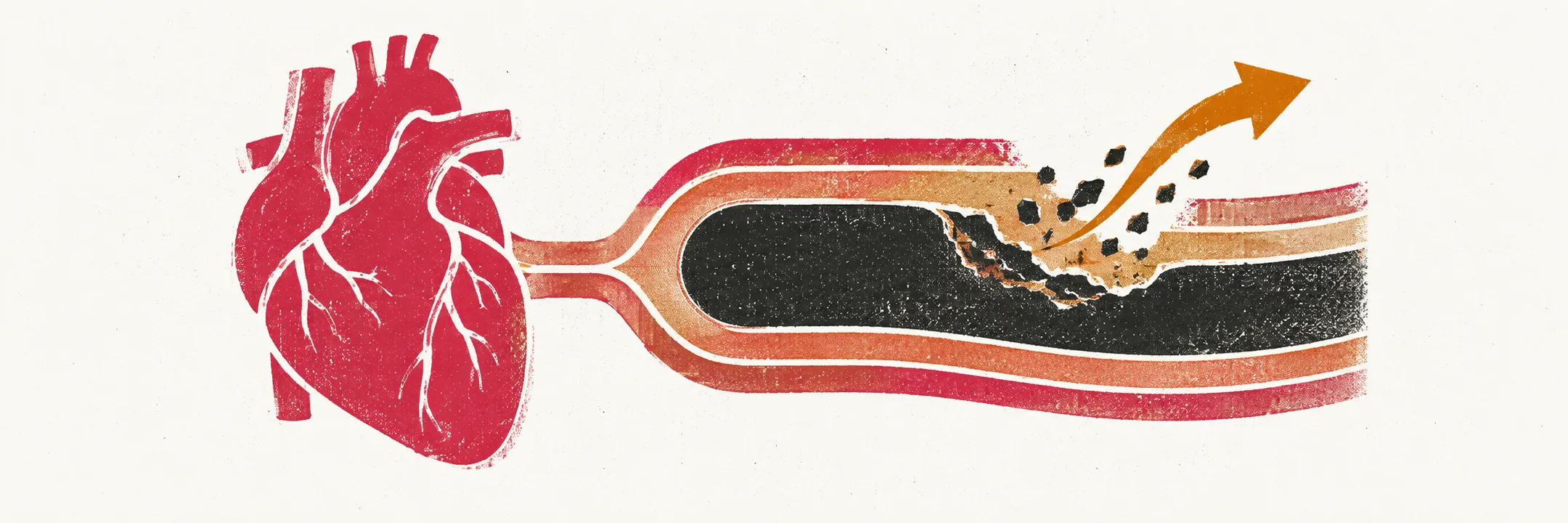
Myocardial infarction is muscle death caused by an interruption of coronary flow, the end point of ischaemia, in which the muscle receives too little blood for its needs. The steps between an unstable plaque and a healed scar decide how much muscle is lost, which complications appear and when, and why the timing of reperfusion runs through the whole topic.
How the artery closes
Atherosclerosis, the build-up of lipid-rich plaque in the arterial wall, is silent until a plaque becomes unstable. A thin fibrous cap over a lipid-rich core ruptures or erodes, platelets adhere to the exposed core and aggregate, and the coagulation cascade lays fibrin over them. The artery narrows or closes, and the muscle it supplies loses its oxygen supply.
Why the damage spreads outward
Ischaemic injury does not appear evenly across the ventricular wall. It begins in the subendocardium, the innermost layer, which sits furthest from the epicardial vessels (the large coronary arteries running over the heart’s surface) and has the least perfusion reserve, and it extends outward toward the epicardium as a wavefront while the artery stays closed. A subendocardial infarct is therefore a smaller version of the same process, and a transmural infarct, one that spans the full thickness of the wall, is the end point of it.
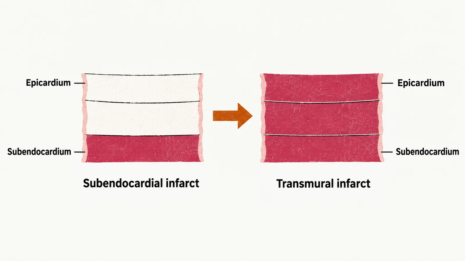
The useful consequence is that the amount of muscle lost is not fixed at the moment of occlusion. It is set by how much was still viable when flow returned, which is why the delay to reperfusion appears in every discussion of outcome.
Reperfusion is also an injury
Reperfusion means restoring flow through the occluded artery. It is better than leaving the artery closed, but it does not only undo damage. The damage inflicted on the myocardium during infarction is the result of ischaemia and of the reperfusion that follows it, and the two are described together as ischaemia-reperfusion injury.
Reperfusion of muscle that has been ischaemic can also fail at the level of the microcirculation rather than the artery. No-reflow means that the epicardial vessel is open and blood still does not reach the tissue, because microvascular obstruction persists: clot and debris have been embolised downstream, reperfusion injury has damaged the capillaries themselves, and individual microvascular susceptibility varies. Angiographic no-reflow is seen in more than 20% of primary angioplasty procedures (reopening the artery by a catheter procedure as the first treatment), although reported rates vary widely with the definition used, and it is associated with a larger infarct, worse left ventricular function and higher mortality. The clinical handling of no-reflow belongs to Reperfusion in Acute Myocardial Infarction.
Stunned and hibernating myocardium
Even when reperfusion succeeds, the muscle it saves does not always start contracting at once. Two forms of dysfunction look like lost muscle and are not.
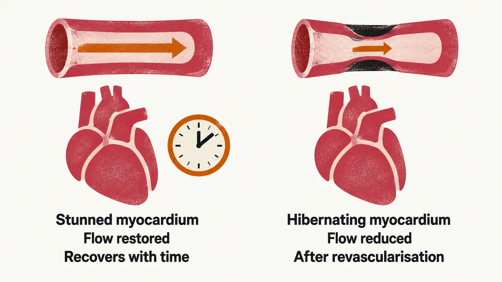
Stunned myocardium is contractile failure that persists after flow has been restored. The cells are alive and the artery is open; recovery takes days to weeks. It is the reason left ventricular function measured immediately after an infarct underestimates the function the patient will eventually have, and the reason decisions about defibrillators are delayed.
Hibernating myocardium is a chronic rather than an acute state: resting flow is persistently reduced, contractility falls to match it, and function recovers after the vessel is revascularised. The distinction matters because only one of the two waits for an intervention: stunned muscle already has its flow back and recovers with time, whereas hibernating muscle recovers only after revascularisation.
How the infarct heals
Muscle that was not saved dies, and the dead muscle is replaced by scar in a sequence that takes weeks and is visible both to the pathologist and to the clinician at the bedside:
| Time after infarction | What is happening in the muscle |
|---|---|
| 4–12 hours | early coagulative necrosis |
| 12–24 hours | neutrophils move into the infarct |
| 1–3 days | nuclei are lost |
| 3–7 days | macrophages clear the dead muscle |
| 7–10 days | granulation tissue forms at the margins |
| 10–14 days | formed granulation tissue with new vessels |
| 2–8 weeks | collagenous scar, spreading from the margins to the centre |
| beyond 2 months | dense scar |
The earliest changes need 6 to 12 hours before they are visible under the microscope, so a heart examined within hours of death can look normal. The sequence also explains why the complications of infarction have a timetable: rupture of the ventricular wall or septum requires dead muscle that has not yet been replaced by scar, so it clusters in the first week, while aneurysm formation and remodelling belong to the weeks and months that follow (see Complications of Myocardial Infarction).
In the first hour, though, this sequence has not yet begun to show, and suspicion of an occluded artery has to come from how the patient presents.