
Diagnosing an acute coronary syndrome (ACS) is a two-step exercise. A 12-lead ECG taken within 10 minutes of first medical contact decides whether the patient belongs to the ST-elevation pathway, which defaults to immediate reperfusion (reopening the blocked artery), or to the non-ST-elevation pathway, where risk stratification sets the timing of angiography. Cardiac troponin, a protein released from injured heart muscle, then establishes whether myocardial injury has occurred at all, which is what separates infarction from ischaemia without necrosis.
Both steps are imperfect on their own. The ECG can be normal in a genuine occlusion, and troponin rises in many conditions that are not atherothrombotic ACS. The diagnosis is made by combining the two with the clinical picture, and by remembering that the initial ECG is a snapshot rather than a verdict.
What the three acute coronary syndromes are
Three entities sit under the ACS umbrella, and the ECG together with cardiac troponin separates them:
- Unstable angina — ischaemia at rest without detectable myocardial injury, so troponin stays normal.
- NSTEMI (non-ST-elevation myocardial infarction) — troponin shows acute myocardial injury, a rise and/or fall with at least one value above the assay’s 99th percentile, but there is no persistent ST-segment elevation.
- STEMI (ST-elevation myocardial infarction) — persistent ST-segment elevation in contiguous leads, generally reflecting an acutely occluded artery, and the only presentation that triggers immediate reperfusion as a default.
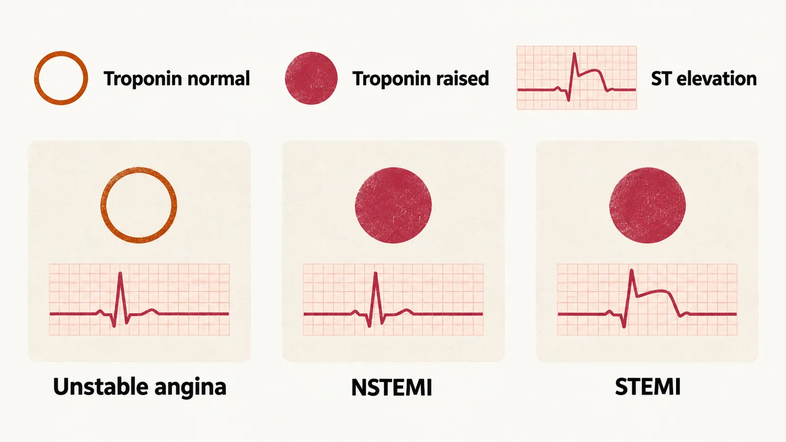
Behind this division sits a pathological picture: total occlusion is paired with transmural infarction, through the full thickness of the wall, and subtotal or intermittent occlusion with subendocardial infarction, confined to the inner layer. That pairing is a useful rule of thumb rather than a law. Collateral flow, prior infarction, multivessel disease and non-atherothrombotic mechanisms all blur the correlation between the surface ECG and the pathological extent of necrosis.
Reading the ECG: thresholds and territory
ST-segment elevation is measured at the J-point, where the QRS complex meets the ST segment, and the threshold depends on lead position and sex:
| Leads | Threshold |
|---|---|
| V2–V3 | ≥2.5 mm in men under 40 years, ≥2 mm in men 40 years or older, ≥1.5 mm in women |
| All other leads | ≥1 mm in any patient |
Two conditions apply to the measurement. First, the elevation has to appear in two contiguous leads — leads that view the same wall, not necessarily neighbours on the paper. Second, the thresholds assume there is no left ventricular hypertrophy (LVH) or bundle branch block, because both produce secondary ST–T changes that mimic injury.
Because each group of leads looks at one wall of the left ventricle, the leads that show the elevation also point to the artery responsible:
| Territory | Leads | Usual culprit |
|---|---|---|
| Anterior (septal to apical) | V1–V4, sometimes V5–V6 | Left anterior descending artery |
| Lateral | I, aVL, often V5–V6 | Circumflex artery or a large diagonal branch |
| Inferior | II, III, aVF | Right coronary artery (RCA) or circumflex |
| Posterior | Tall R and ST depression in V1–V2 | RCA or circumflex, via posterior branches |
Three details of the lead pattern are worth keeping. In inferior infarction, comparing the height of the elevation in lead III with lead II points to the culprit: elevation that is taller in III suggests the RCA, and taller in II suggests the circumflex, because the two leads look at the inferior wall from different sides. Because RCA occlusion can involve the right ventricle, right-sided leads (V3R–V4R) are recorded in inferior infarction, and right ventricular involvement changes management toward volume support rather than diuresis. Posterior infarction is the pattern that is regularly missed, because the standard leads look at it from the front: a dominant R wave with ST depression in V1–V2 is the mirror image of an elevation recorded from behind, and posterior leads V7–V9 confirm it.
ST depression is not automatically a non-ST-elevation event. Depression in one territory can be the reciprocal reflection of an elevation in another, because one injury current directed toward one wall is directed away from the opposite wall. Anterior elevation produces inferior depression; inferior elevation produces high-lateral or anterior depression. When depression appears without an obvious explanation, the next move is to look for elevation elsewhere rather than to close the case.
Two further ECG patterns carry disproportionate weight. ST elevation in lead aVR with widespread ST depression suggests left main or severe three-vessel disease and belongs in the immediate-angiography group, even though the criteria for ST-elevation infarction are not met. A new left bundle branch block, by contrast, no longer carries that implication.
When left bundle branch block is present
Left bundle branch block (LBBB) distorts repolarisation and produces ST–T changes discordant to the QRS complex, pointing in the opposite direction to it, so the ordinary ST-elevation criteria cannot be applied when the QRS is wide. Two things follow from that.
First, a new LBBB is not a trigger by itself. The 2023 European guideline notes that LBBB prevents an accurate assessment of whether ST elevation is present, and that a patient with this pattern together with signs or symptoms highly suspicious for ongoing ischaemia should be managed like a patient with clear ST elevation. The 2025 American guideline says that new or presumably new LBBB should not be considered diagnostic of acute myocardial infarction in isolation, and that in an asymptomatic patient it does not constitute a STEMI equivalent.
So the clinical picture, not the conduction abnormality, decides the pathway. A patient with LBBB and no ongoing ischaemic symptoms follows the non-ST-elevation pathway, where troponin, symptoms and haemodynamic state set the timing of angiography. A patient with LBBB plus signs or symptoms highly suspicious for ongoing ischaemia is managed in the same way as a patient with clear ST elevation, and instability such as shock, refractory pain or a life-threatening arrhythmia makes that call urgent.
Second, occlusion can still be recognised inside an LBBB by using criteria designed for it. The original Sgarbossa criteria looked for concordant changes, running in the same direction as the QRS — ST elevation ≥1 mm in a lead where the QRS is positive, or ST depression ≥1 mm in V1–V3 — and for discordant ST elevation of ≥5 mm, and a weighted score of 3 or more was highly specific. The weakness was sensitivity: most occlusions were missed. The modified Sgarbossa rule replaces the 5 mm absolute criterion with a ratio, treating discordant ST elevation as abnormal when it is at least 25% of the preceding S-wave depth. The modified rule performs better than the original in the small retrospective series that introduced it, but the confidence intervals are wide, so these criteria support a diagnosis rather than settle it. Clinical assessment and serial troponin remain part of the decision.
Troponin and the diagnostic algorithm
Cardiac troponin I or T is the biomarker of choice. Myocardial injury is defined as a rise and/or fall of troponin with at least one value above the assay’s sex-specific 99th percentile upper reference limit, the level exceeded by only the top 1% of a healthy reference population; a single static elevation cannot distinguish acute injury from chronic elevation. Adding clinical evidence of ischaemia — symptoms, new ECG changes, new wall motion abnormality, or a coronary thrombus on imaging — converts injury into infarction.
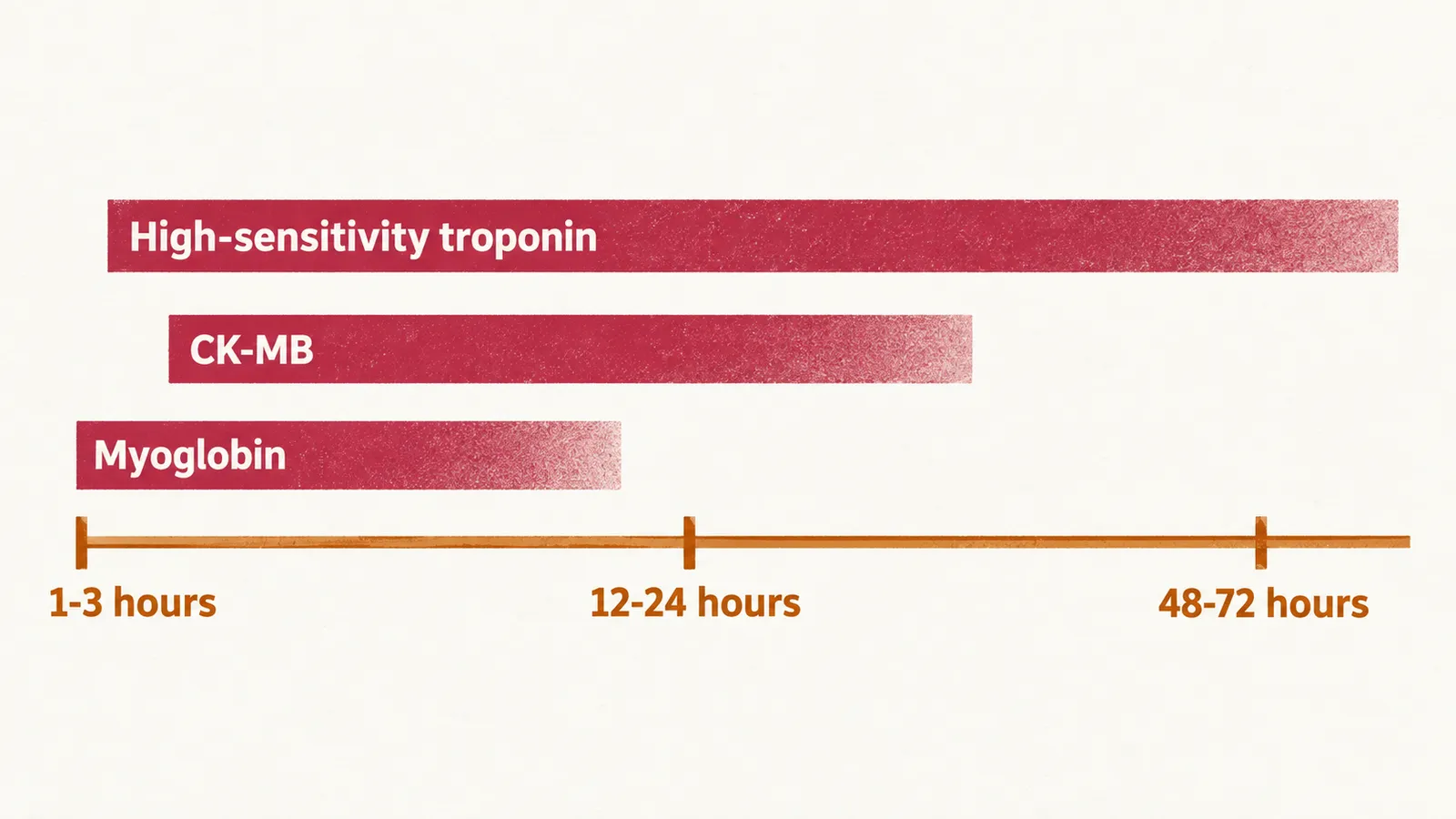
Timing and magnitude differ by marker, which is why older markers still appear in teaching:
- High-sensitivity troponin: detectable within 1–3 hours of symptom onset, useful for rapid rule-in and rule-out, and it remains elevated for days to about two weeks.
- CK-MB: rises over 4–6 hours, peaks around 12–24 hours, normalises within 48–72 hours, which once made it useful for detecting reinfarction.
- Myoglobin: rises earliest but is not cardiac-specific, so it has been abandoned in practice.
For patients without ST elevation, current European guidance recommends the 0/1-hour algorithm using high-sensitivity troponin as the preferred rule-in/rule-out strategy: troponin is measured at presentation and again 1 hour later. A 0/2-hour algorithm is an accepted alternative and the older 0/3-hour protocol a further option. That last protocol requires freedom from pain and a low-risk GRACE score, a score that forecasts death and reinfarction. The algorithm assigns most patients at the first reassessment, leaving the remainder for further observation and testing. The exact cut-offs are assay-specific, which is why each laboratory implements the version validated for its own platform.
Echocardiography in the acute phase
Echocardiography carries part of the diagnostic load that the ECG and troponin leave. A new regional wall motion abnormality is objective evidence that the injury is ischaemic, and it completes the definition of infarction when the ECG is inconclusive. The same study shows how much muscle is not contracting and what the left ventricular function is, which later treatment decisions depend on; it identifies the mechanical complications described in Complications of Myocardial Infarction; and it turns up competing explanations such as a pericardial effusion, aortic dissection or significant valve disease.
Risk scores
Once the diagnosis is made, the next question is how dangerous the event is. Two scores appear throughout the ACS literature, and both forecast rather than diagnose. GRACE 2.0 predicts death and the combined outcome of death or myocardial infarction, both during the acute admission and at one year; a score above 140 places the patient in the high-risk group whose angiography is considered early. TIMI publishes separate scores for unstable angina or NSTEMI and for STEMI. Both help weigh how intensive treatment should be, neither makes the diagnosis, and routine use of risk scores has not been shown to reduce cardiovascular events.
How myocardial infarction is classified now
Confirming an infarction leaves one more question: what caused it in the artery. Until 2026, infarction was classified by number: type 1 for spontaneous atherothrombotic events, type 2 for supply–demand imbalance, type 3 for death before biomarkers could be obtained, types 4a to 4c for procedure-related infarction, stent thrombosis and restenosis, and type 5 for coronary artery bypass grafting. The Fifth Universal Definition of Myocardial Infarction, published in August 2026, replaces that enumeration with three categories that describe what is happening in the coronary artery:
- Primary MI — spontaneous, caused by an acute coronary pathology. Atherothrombotic plaque rupture or erosion is the most common, but the category also covers spontaneous coronary artery dissection, coronary embolism, vasospasm, and restenosis, stent thrombosis or graft failure occurring more than 30 days after a procedure.
- Secondary MI — myocardial oxygen supply–demand imbalance caused by another acute condition. The diagnosis is confirmed when the ischaemia resulting from that mismatch is a consequence of obstructive coronary disease without evidence of an acute coronary pathology, or when the resulting injury is enough to cause a new regional wall motion abnormality or loss of myocardial viability.
- Procedure-related MI — acute ischaemia and injury arising from a complication of a percutaneous or surgical cardiac procedure within 30 days. The criteria are identical for both routes and they are not biomarker thresholds: what defines the event is the coronary complication. When the complication arises during the index procedure, or during treatment of an acute myocardial infarction, both features are required — a coronary complication and a new or presumed-new regional wall motion abnormality or loss of viability.
The older numbers are still used in textbooks, trials and everyday clinical speech, so they remain worth knowing. The practical difference is treatment: primary MI follows the antithrombotic and catheterisation pathway, whereas secondary MI is treated by correcting the condition that created the mismatch, and procedure-related MI is managed as a procedural complication. Documentation now records the responsible mechanism — atherothrombosis, dissection, embolism, vasospasm, restenosis, stent thrombosis or graft failure — so naming it is part of the diagnosis.
Telling primary, secondary and procedure-related MI apart at the bedside
Because the categories differ in treatment, the bedside question is which one a raised troponin belongs to. The category is decided by what the troponin result sits alongside, not by its height.
- A patient with chronic kidney disease often has a raised troponin at baseline, so acute injury requires a rise and/or fall rather than a single elevated value.
- Sepsis, tachyarrhythmia, severe anaemia and other acute illnesses produce supply–demand mismatch; secondary MI is the diagnosis when obstructive coronary disease is present and there is no acute coronary pathology, and when the injury has caused a new wall motion abnormality or loss of viability.
- A troponin rise after a percutaneous or surgical procedure counts as infarction only when there is a coronary complication, and only within 30 days of the procedure.
- A spontaneous event with an acute coronary pathology is primary MI, whichever mechanism is responsible; a dissection should be considered in women under 50 and in pregnancy or after delivery.