
The coagulation factors are the clotting proteins, numbered with Roman numerals (FII, FV, FVIII and so on), that act in sequence to form a clot. The coagulation factor deficiencies are a group of bleeding disorders whose phenotype depends on how much factor activity remains. The bleeding they cause collects in joints, muscles and closed spaces, and the pathway involved can be read from the prothrombin time (PT) and the activated partial thromboplastin time (aPTT).
The bleeding pattern
Patients with coagulation factor deficiencies experience recurrent episodes of bleeding into the joints, muscles and closed spaces, either spontaneously or due to preceding trauma. This deep pattern is what separates them from the surface bleeding of platelet and vessel disorders.
As a general rule, the phenotype of bleeding in coagulation disorders is related to the amount of activity of the coagulation factor.
The inherited deficiencies
The most common form of the coagulation deficiencies is haemophilia, X-linked diseases that affect FVIII and FIX, which are haemophilia A and haemophilia B, respectively.
Other forms of coagulation disorder, rare in comparison to haemophilia, are mostly autosomal recessive disorders related to FII, FV, FVII, FX, FXI and FXIII. FXII deficiency does not cause a bleeding phenotype, perhaps because there is a redundant pathway of activation for FXI, which is activated by the activated form of FII that is made by the extrinsic pathway.
Laboratory testing
The main laboratory tests that can be used for the diagnosis of coagulation factor-related disorders are:
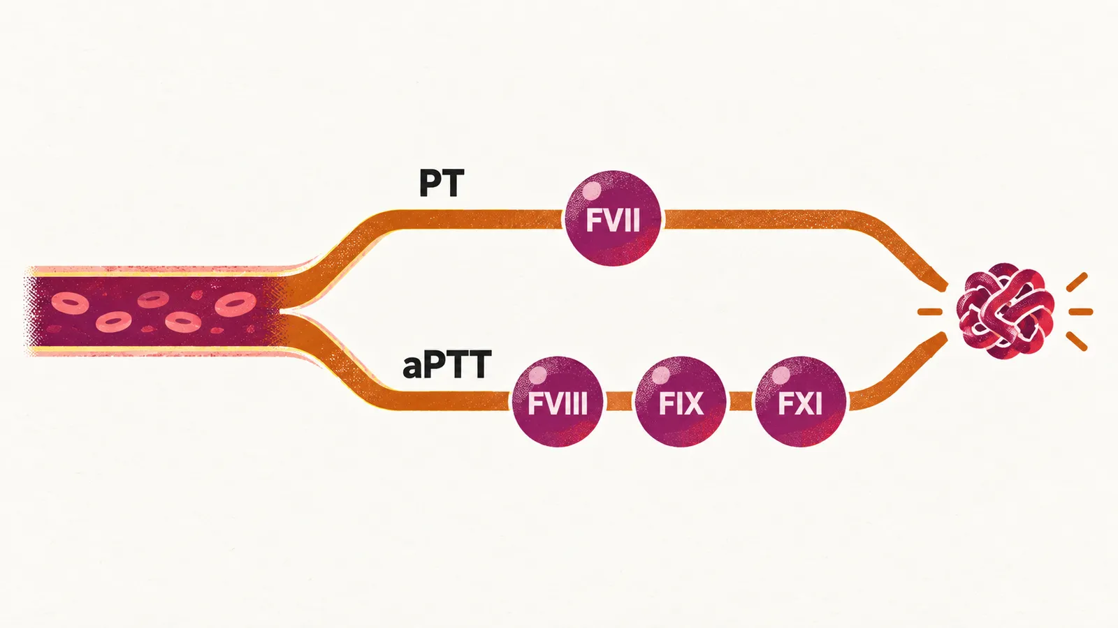
- PT — for the extrinsic pathway; a prolonged PT indicates deficiency of FVII
- aPTT — for the intrinsic pathway; a prolonged aPTT indicates deficiency related to FVIII, FIX or FXI
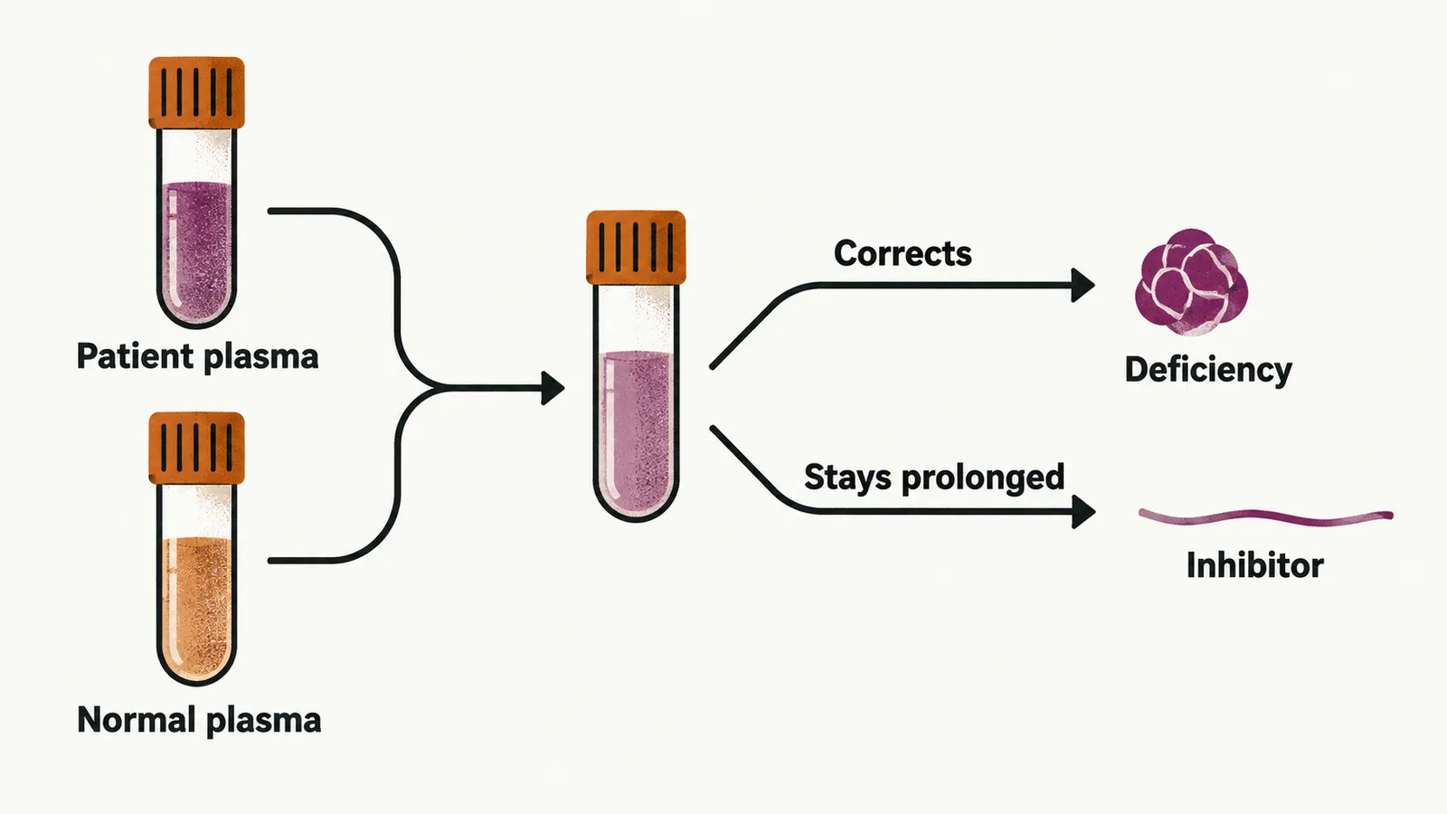
- mixing test — the patient’s plasma is mixed with normal plasma; correction of the prolonged PT or aPTT indicates a factor deficiency, while failure to correct indicates an inhibitor, and the test is also used to detect an anticoagulant
The mixing test works because normal plasma supplies the factor that is missing. When a factor is deficient, adding normal plasma restores the pathway and the clotting time corrects; when an inhibitor is present instead, the inhibitor also acts on the factor supplied by the normal plasma, so the time stays prolonged.
Acquired deficiencies
Acquired deficiencies of coagulation factors are more common in comparison to the inherited ones. They arise in four settings:
- haemorrhagic diathesis of liver disease
- disseminated intravascular coagulation (DIC)
- vitamin K deficiency
- development of alloantibodies to coagulation factors in patients with a deficiency who need infusion of these factors after episodes of bleeding
The alloantibodies form against the infused factor in patients who already have a deficiency, and haemophilia is the most common of the inherited deficiencies.