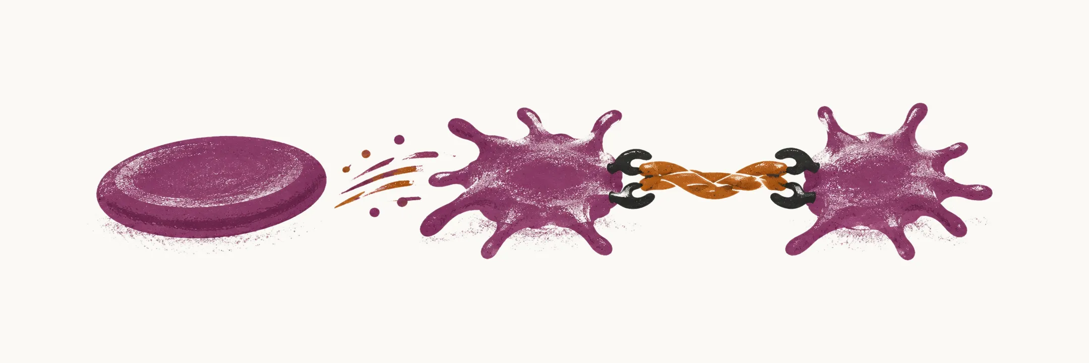
Platelets are small, nucleus-free cell fragments that circulate in the blood. Together with the blood vessel wall, the coagulation pathway (the sequence of clotting factors that forms a clot) and the fibrinolytic pathway (which dissolves clots) they make up the four determinants of haemostasis, the process that keeps blood fluid in intact vessels and seals a breach. A bleeding disorder that arises in the platelets follows from either their number or the way they work.
Normal platelet production and survival
The normal count of platelets in the blood is about 150,000 to 450,000 per microL (per microlitre), and it is mainly under the control of thrombopoietin, a hormone that is made by the liver. Thrombopoietin is produced at a steady rate and is removed from the circulation by platelets and megakaryocytes, the bone marrow cells that produce platelets, so when the platelet count falls, more of the hormone remains free to act on the bone marrow and drive it to make more platelets.
The average life span of a platelet is about 7 to 10 days. Platelet synthesis can be increased several-fold in response to inflammation, especially in response to IL-6, an inflammatory signalling protein (interleukin-6) that raises thrombopoietin production.
Platelets are also held in reserve. About one-third of the total platelet mass is located in the spleen. This pool is held aside rather than lost, and it grows when the spleen enlarges; in marked splenomegaly the sequestered fraction is large enough that the number of platelets in the blood can fall to less than 40,000 per microL.
How platelets are activated
In an intact vessel, endothelial cells keep platelets in a resting, anti-thrombotic state so that they do not adhere or aggregate where they should not. Platelets are drawn out of this state by the exposure of collagen and von Willebrand factor (VWF) in the sub-endothelium, by the shear stress of flowing blood, and by an inflammatory state of the endothelium.
When the endothelial cells become activated, their effect changes to a thrombotic one: the platelets attach to them, which causes activation of some intracellular pathways in the platelets and leads to activation of the surface glycoprotein Gp IIb/IIIa. Once Gp IIb/IIIa is active, it binds fibrinogen and links platelets to one another, which is platelet aggregation at the site of activation. The same activation drives degranulation, the release of nucleotides, procoagulants, growth factors and adhesive proteins that recruit further platelets and support clot formation.
Mediators that act on the vessel wall also act on platelets: nitric oxide, which makes the vessel dilate, inhibits aggregation, while endothelin, which makes it constrict, activates aggregation.
The main groups of platelet disorder
Because a platelet problem lies either in the number of platelets or in how they work, the forms of platelet disorder that are clinically the most relevant and frequent fall into four groups:
- thrombocytopenia — too few platelets (see Platelet Count Disorders: Thrombocytopenia and Thrombocytosis)
- the thrombotic thrombocytopenic microangiopathies (TTM), in which small-vessel thrombosis and platelet consumption occur together with microangiopathic haemolytic anaemia (see Thrombotic Thrombocytopenic Microangiopathies: TTP and HUS)
- thrombocytosis — too many platelets (see Platelet Count Disorders: Thrombocytopenia and Thrombocytosis)
- qualitative disorders of platelet function, in which the count is normal but the platelets do not work properly