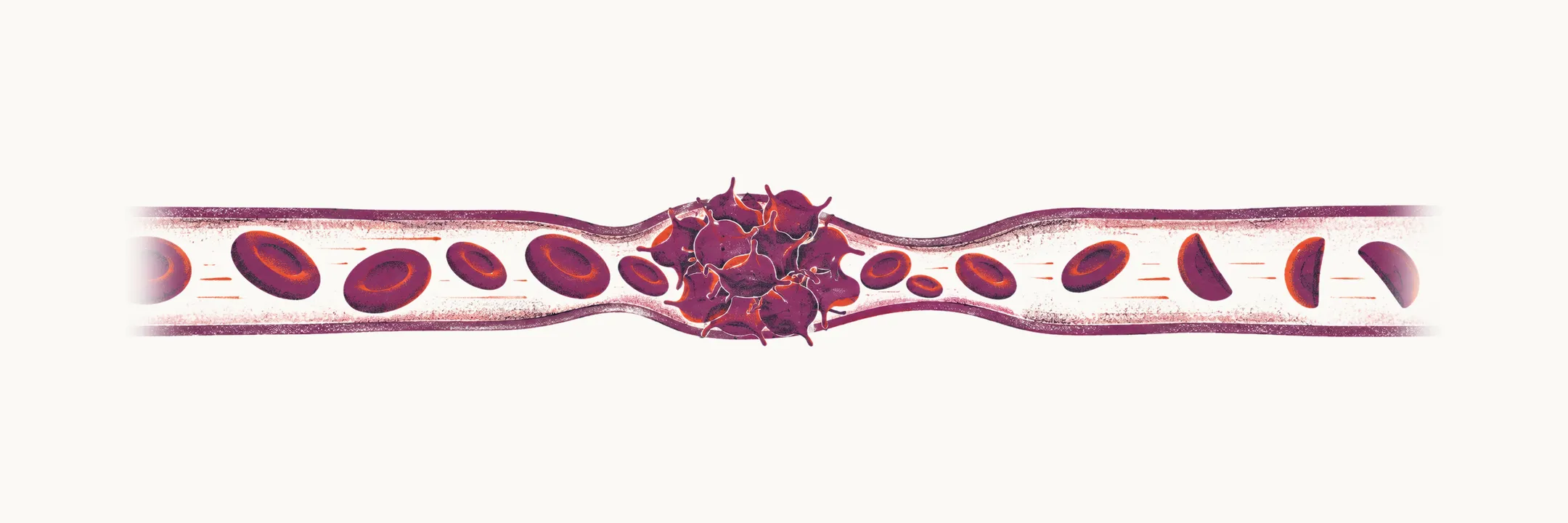
In the thrombotic thrombocytopenic microangiopathies, small-vessel thrombosis and platelet consumption occur together with microangiopathic haemolytic anaemia, so a patient has a low platelet count and haemolysis at the same time.
The group
Thrombotic thrombocytopenic microangiopathies (TTM) are a group of disorders characterised by:
- microangiopathic haemolytic anaemia, or MAHA, defined by fragmented red cells, or schistocytes, together with evidence of haemolysis
- thrombocytopenia
The two features are linked by a single process: platelet-rich thrombi form in the small vessels, and the red cells forced through them are physically sheared and break apart, which is why fragments appear on the smear. The findings in TTM are:
- evidence of haemolysis: increased LDH (lactate dehydrogenase), increased unconjugated bilirubin and decreased haptoglobin
- microvascular thrombosis
The disorders which are included in the group are:
- thrombotic thrombocytopenic purpura, or TTP
- haemolytic-uraemic syndrome, or HUS
- syndromes complicating bone marrow transplants
- some infections
- pregnancy
- vasculitis
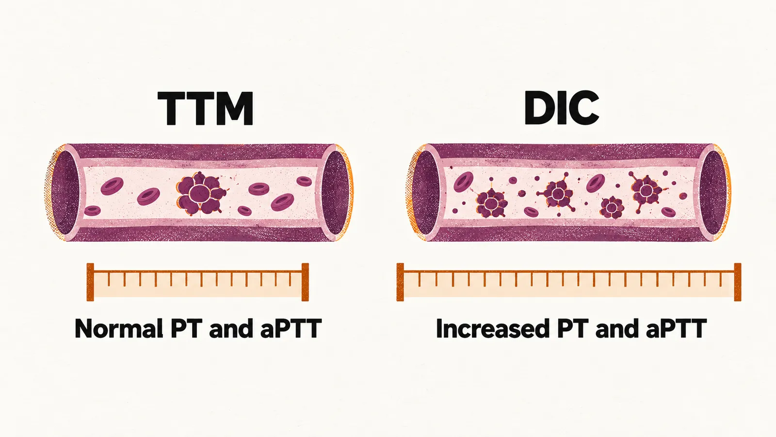
Disseminated intravascular coagulation (DIC) produces both the microangiopathy and thrombocytopenia, so it resembles the TTM. The distinguishing factor between DIC and TTM is the decreased level of coagulation factors in DIC, which leads to an increased prothrombin time (PT) and activated partial thromboplastin time (aPTT); these are in the normal range in all the TTM conditions. This is the differential diagnosis between DIC and TTP.
Haemolytic-uraemic syndrome
Two forms of HUS are seen:
| Form | Cause | Course | Mortality |
|---|---|---|---|
| typical HUS | mostly related to infection with E. coli O157:H7 | acute, after a diarrhoeal illness | less than 5% |
| atypical HUS, or aHUS | mostly due to genetic defects in complement genes or antibodies against them | over a chronic time course | about 26% |
Typical HUS is managed with supportive care, including fluids, electrolytes and renal replacement when needed. Atypical HUS is driven by uncontrolled complement activation, so it is treated with a complement inhibitor such as eculizumab, a C5 antibody that blocks the pathway producing the thrombosis.
Thrombotic thrombocytopenic purpura
TTP was first described by Moschcowitz as a pentad of:
- microangiopathic haemolytic anaemia, or MAHA
- thrombocytopenia
- renal failure
- neurologic findings
- fever
The full pentad is not present in every patient, but the combination of MAHA and thrombocytopenia without another cause is what should raise the diagnosis. TTP is more common in HIV-positive patients and in pregnant women.
Pathogenesis
The pathogenesis of TTP has two forms:
- the inherited form, called Upshaw-Schulman syndrome — a genetic deficiency in the production of ADAMTS13, which cleaves the macromolecules of VWF (von Willebrand factor, the protein that tethers platelets to the injured vessel wall). A defect of ADAMTS13 alone is not able to produce the TTP condition, because in these patients TTP occurs episodically and mostly during pregnancy.
- the acquired form, also called idiopathic TTP or ITTP — it is related to the production of antibodies against ADAMTS13. The most common cause of TTP is the immune-related one. ITTP is more common in women than in men.
Diagnosis and treatment
The test that confirms the diagnosis is the assay for the activity of ADAMTS13: TTP is supported when it is less than 10% of the normal range of activity.
Therapeutic plasma exchange, or TPE, is the main treatment for patients with TTP; it is continued until the platelet count is normal and the signs of haemolysis have resolved for at least 2 days. In addition to TPE, rituximab and caplacizumab, an anti-VWF nanobody, can be used, but they are indicated only for patients with a diagnostic ADAMTS13 level lower than 10% of activity. Even with treatment, recurrence is common — reported in roughly 20-50% of patients who survive the initial episode — so ADAMTS13 activity is followed after remission.
When Moschcowitz first described TTP, the mortality rate was about 85-100%, and nowadays it is about 10-30%, thanks to early diagnosis before the occurrence of the full-blown syndrome and to plasma replacement in these patients.
TTP thus turns on ADAMTS13, the enzyme that acts on von Willebrand factor. When von Willebrand factor itself is deficient or defective, the result is a bleeding disorder rather than thrombosis: von Willebrand disease.