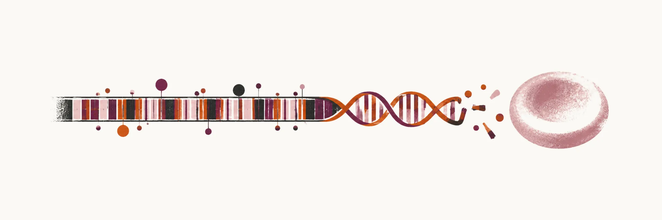
β-thalassemia is the form of thalassemia, an inherited reduction in the synthesis of a globin chain, in which the synthesis of the β-globin chain is reduced or absent. It is also known as Mediterranean thalassemia, because of its high prevalence in Italy and Greece.
The two kinds of β-globin allele
Two kinds of β-globin allele cause the disease:
- β0 is a completely non-functional gene, so no β-globin is produced at all
- β+ is a slightly non-functional gene, so β-globin is produced in reduced amount
The mutations, which are mostly point mutations, can be a mutation in the promoter, a mutation at the exon-intron junction that disturbs mRNA splicing, the introduction of a new splicing site, a 3’-end mutation that prevents polyadenylation of the mRNA, a mutation that prevents translation initiation, or a premature stop codon.
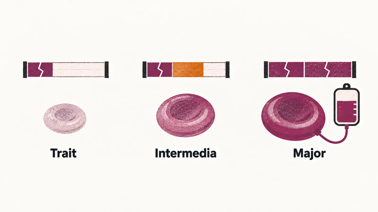
If one of the alleles is mutated, the form is called the trait or minor; if both alleles are mutated, it is called the major form. One phenomenon shapes both: most of the red cells made in the bone marrow die very early, inside the bone marrow itself. This is called ineffective erythropoiesis, and it is the main reason for the anemia of β-thalassemia.
Forms of β-thalassemia
The forms differ in how much β-globin is made and in how much transfusion the patient needs.
Beta thalassemia minor or trait
The trait is heterozygous, with one mutated allele. Its laboratory findings are a microcytic hypochromic anemia with an increased HbA2 and HbF, and a decreased HbA.
Beta thalassemia intermedia or transfusion-independent
Intermedia is the middle form, and it is defined in two ways that fit together: there is at least one β+ mutation, so there is a low-grade production of β-globin in the erythroid cells, and the patient has anemia but does not need transfusion.
The pathophysiology of β-thalassemia intermedia is tightly related to the amount of unpaired α-globin chain, so anything that reduces that unpaired α — or increases the γ chains that can pair with it — makes the disease milder. The disorders that are counted as β-thalassemia intermedia are:
- homozygosity for the β+ allele
- heterozygosity for β+ and β0
- co-inheritance of β-thalassemia genes with mutated genes for increased γ chain synthesis, such as hereditary persistence of fetal hemoglobin, or HPFH
- co-inheritance of a homozygous β+ gene with an α-thalassemia deletion
- co-inheritance of a heterozygous β-thalassemia gene with triplicated or quadruplicated α genes
Beta thalassemia major or transfusion-dependent
The clinical manifestation of β-thalassemia major shows itself during 2 to 6 months of age, once the fetal hemoglobin of infancy has fallen and the missing β chains begin to matter. The early clinical signs are pallor, growth retardation, jaundice, gallstone, hepatomegaly, splenomegaly, and a skeletal change mainly in the face, which is called the chipmunk face.
The late clinical signs are:
- endocrine signs due to iron overload — hypogonadism, diabetes mellitus, hypothyroidism
- cardiac signs due to iron overload, such as heart failure
- a hyper-coagulable state
- pulmonary hypertension
- aplastic crisis due to parvovirus B19 infection
Laboratory findings and complications of the severe forms
In the more severe forms of β-thalassemia — anything other than the trait — the laboratory shows:
- microcytic hypochromic anemia
- hemolytic anemia
- increased HbA2 and HbF
- decreased HbA
- increased iron storage, with an increased ferritin
- reticulocytosis
- anisocytosis
- poikilocytosis
In the same severe forms, the complications fall into groups:
- complications related to hemolysis — hepatomegaly, splenomegaly, hyperbilirubinemia and gallstone
- skeletal change due to expansion of the hematopoietic bone marrow — the chipmunk face
- complications related to iron overload — these affect mainly the endocrine system and the heart, causing hypothyroidism, diabetes mellitus, liver diseases, heart failure and arrhythmia, and they occur in the transfusion-independent form too, because of the increase in gastric absorption of iron
- complications related to transfusion, such as probable infections
- a hyper-coagulable state
- pulmonary hypertension
Treatment of beta thalassemia
The treatments answer the problems above: anemia and iron overload are handled with transfusion and chelation, ineffective erythropoiesis with luspatercept, and the underlying gene defect with transplantation or gene therapy.
Transfusion and iron chelating agents. The goal of transfusion is to reach a Hb of 9-10.5 g/dl after each transfusion, which is about every 2 to 4 weeks. Iron chelating agents are important for preventing the complications related to iron overload, which come both from the increase in gastric absorption and from blood transfusion, and which can cause cardiomyopathies and endocrinopathy. The chelating drugs are deferasirox and deferiprone by mouth, and deferoxamine intravenously.
HSCT (hematopoietic stem cell transplantation). It gives a cure rate of more than 80% when the hematopoietic stem cells come from a sibling donor.
Improving ineffective erythropoiesis. Luspatercept is used for this.
Gene therapy. It uses a modified HbA gene that is transported to the patient by lentiviruses that infect the CD34+ hematopoietic stem cells. The product that does this received a marketing authorisation in Europe, but the authorisation was later withdrawn at the request of the company for commercial reasons.
Causes of death
Despite treatment, the main causes of death in β-thalassemia are still related to iron overload in late life, mainly heart failure, although this has decreased a little with the use of iron chelator agents. One cause that increased during the last decades was death due to infection after transfusions, such as HBV, HCV and HIV.
The other type of thalassemia, α-thalassemia, arises from deletion of α-globin genes rather than point mutation of the β-globin gene, and its severity follows the number of genes deleted.