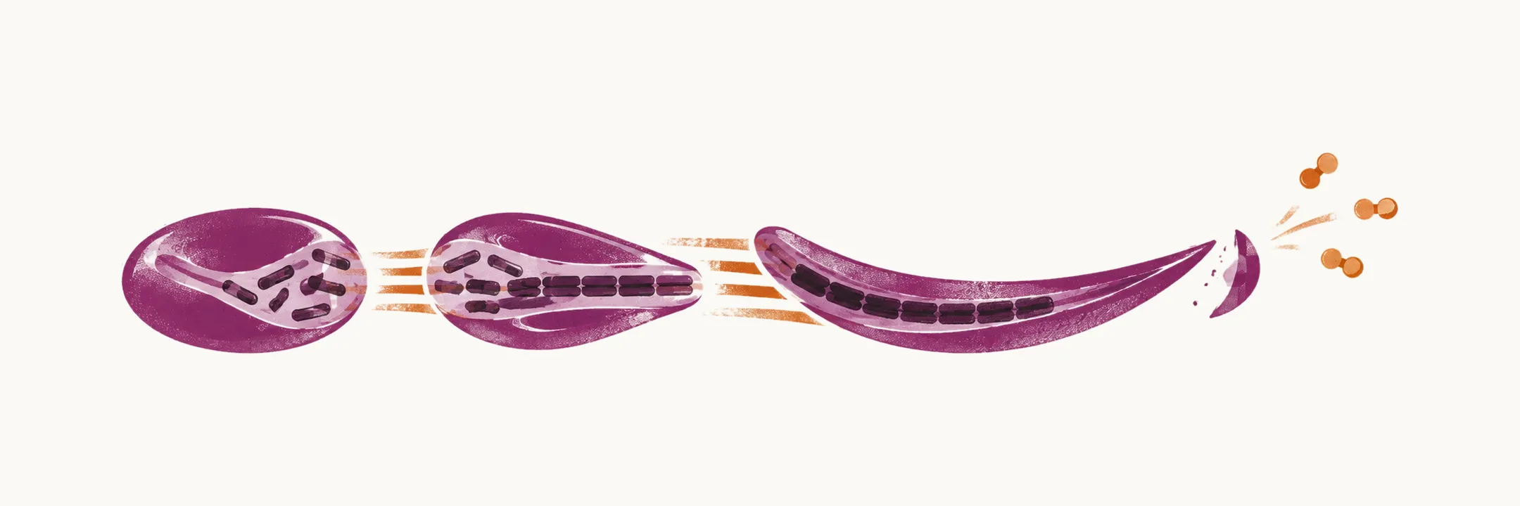
The most common familial inherited hemolytic anemia, an inherited anemia in which red cells are destroyed early, is sickle cell disease, or SCD. It comes from a single amino acid substitution in the β-globin gene, the gene for the β chain of hemoglobin, and the clinical picture grows out of what that substitution does to the hemoglobin molecule.
The mutation and the genotypes it produces
The mutation replaces the 6th glutamic acid of the β-globin chain with valine. The hemoglobin it produces, HbS, is the molecule behind every form of the disease; which form a patient has depends on what the second β-globin allele carries.
- Homozygous HbSS has the valine substitution on both alleles. This is the most prevalent form of sickle cell disease and the most severe. HbA is completely replaced by HbS.
- Sickle cell trait (HbAS) has the substitution on one allele and a normal allele on the other. About half the hemoglobin is HbS and the other half is HbA.
- HbSC has both genes mutated: one carries the HbS mutation, the other the replacement of the 6th glutamic acid with lysine, which is the HbC mutation. About half the hemoglobin is HbS and half is HbC, and no HbA is made.
Sickle cell trait and HbSC are completely different from one another. In sickle cell trait a person carries one mutated gene with a normal gene beside it, whereas in HbSC both genes are mutated. Sickle cell trait is carried by roughly 1 in 12 African Americans. Two things are important in sickle cell trait. The first is monitoring, to prevent an HbSS offspring. The second is the high chance of exertional rhabdomyolysis during exercise, which can cause sudden death in these patients.
Normally in the adult the distribution of the different forms of hemoglobin is 96% HbA, 3% HbA2 and 1% HbF. In sickle cell disease that distribution changes because of the β-globin mutations: in homozygote sickle cell disease HbA is completely replaced by HbS, and in HbSC the HbA is replaced by a mixture of HbS and HbC.
Where the disease is common
Sickle cell disease is believed to have arisen 7000 to 22000 years ago, because the heterozygote, the HbS-HbA phenotype (sickle cell trait), has a superior fitness and an intrinsic resistance to malaria infection. The prevalence of this form of hemoglobin is therefore higher in regions with more malaria infection:
- tropical and sub-tropical regions
- Africa
- the Middle East
- Mediterranean regions
- India
Why the red cell sickles
The manifestation of sickle cell disease is masked until the 5th to 6th month of age, because of the HbF in the red cells of the child, which is present at a high fraction until that age. HbF has no affinity for polymerisation, so while it is present in quantity the red cell is protected. After that age HbF is reduced physiologically, the proportion and concentration of HbS in the cell increase, and the disease begins.
The main mechanism of the disorder is the high tendency of HbS to polymerise, that is, to link into polymers, in the deoxygenated state. HbA and HbF do not share this tendency, so the polymer forms only from HbS.
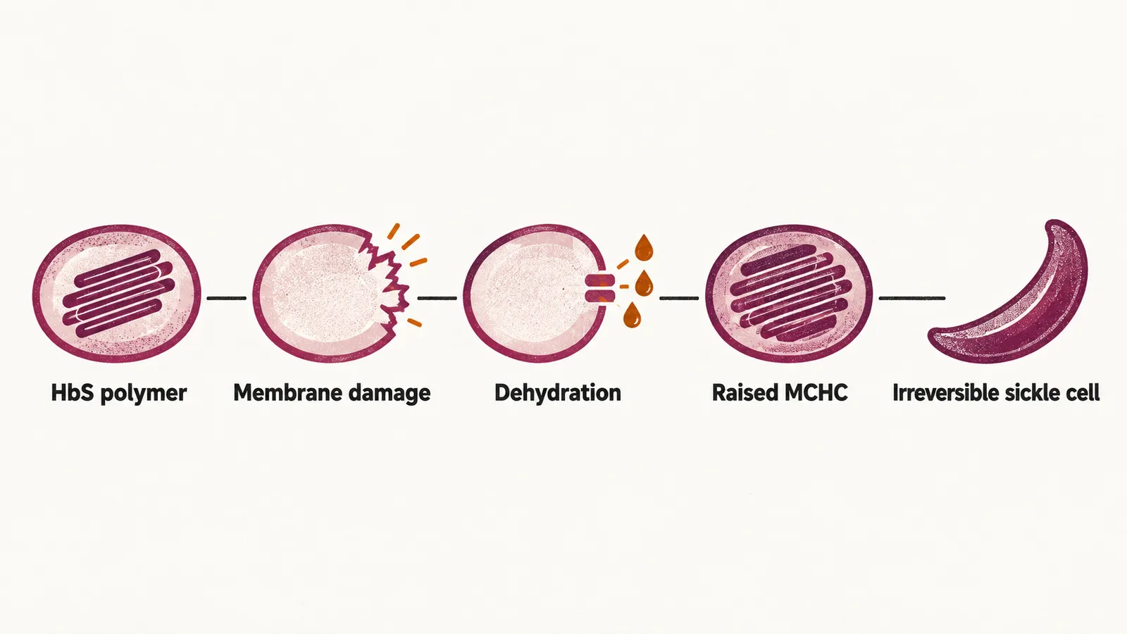
At first the sickle cells that form are reversible: reoxygenation dissolves the polymer and the cell returns to its normal shape. With time, and with the cumulative damage they do to the red cells, the sickle form becomes irreversible. These permanently deformed cells are called the irreversible sickle cells, or ISCs, and they are prone to hemolysis. The damage accumulates step by step:
- the hypoxic polymer of HbS penetrates the red cell membrane, which causes vesiculation and the release of micro-particles from the cell
- the activity of the K/Cl channel and Psickle increases, so potassium and water leave the cell and it becomes dehydrated
- the dehydration raises the MCHC, the mean corpuscular hemoglobin concentration, that is, the concentration of hemoglobin in the red cells, and a raised MCHC makes HbS polymerisation more likely, so this step feeds the process that caused it
- phosphatidylserine moves from the inner to the outer surface of the membrane, which increases oxidative stress in the cell
- at the end, ISCs are formed, deformed for good
What decides whether sickling matters clinically
Three factors determine how much clinically important red cell sickling occurs.
The first is the level of hemoglobin other than HbS in the red cell. The higher the proportion of HbS, the higher the chance of polymerisation. In the homozygote form almost all the hemoglobin is HbS. In the HbSC form about half is HbS and the rest is HbC, which does not take part in the formation of sickle cells. In sickle cell trait almost half is HbS and the other half is HbA, which reduces the chance of sickle formation.
The second is the concentration of HbS in the red cell. A dehydrated cell has a raised MCHC, so in any form of sickle cell disease dehydration raises the chance of sickle formation. In α-thalassemia, an inherited reduction of α-globin synthesis, by contrast, the concentration of hemoglobin in the cell is very low, so a person who has α-thalassemia beside sickle cell disease sickles less.
The third is the passage velocity of the red cells through the microvasculature, the smallest vessels. The faster the cells pass, the lower the chance of a vaso-occlusive episode, a blockage of small vessels by sickled cells, and of sickle formation. Anything that slows that passage, such as inflammation, raises the chance of sickling and vaso-occlusion.