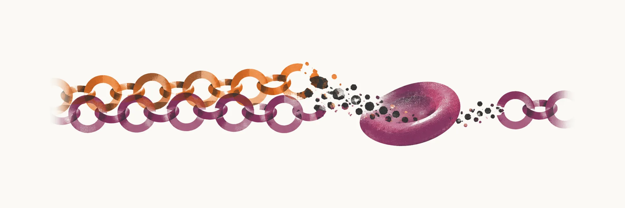
Thalassemia is an inherited autosomal recessive disease that causes a decrease in the synthesis of the α- or β-globin chains, the two kinds of chain that pair to build adult hemoglobin. Which chain is affected decides which of the two forms a patient has: α-thalassemia involves the α-globin or HBA gene, and β-thalassemia involves the β-globin or HBB gene.
Why an imbalance damages the red cell
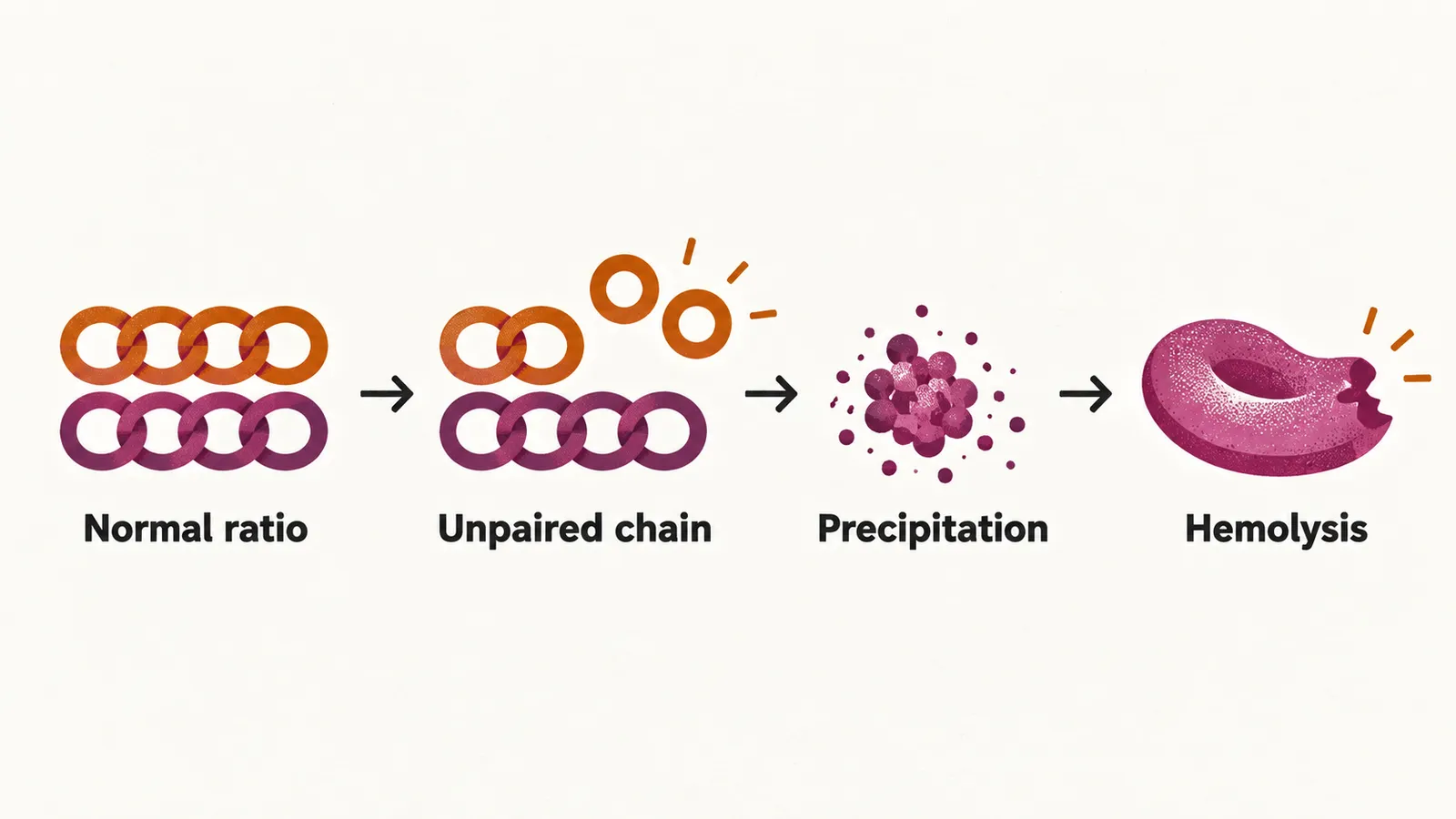
In normal subjects the ratio of production of the α and β globin chains is tightly under control, at about 1±0.05. When the synthesis of one chain falls, that balance is lost and the normal chain is left in excess, unpaired. Two consequences follow, and together they are the basis of the pathophysiology of thalassemia:
- low production of normal hemoglobin, because one of the chains needed to build it is in short supply
- precipitation of the excess chain, which damages the red cells and causes hemolysis, the destruction of red cells
Genetic basis
Thalassemia is caused by a deletion or a point mutation of a globin gene, and which of the two dominates differs between the types:
- in α-thalassemia there is mainly deletion of one or more α-globin genes on chromosome 16
- in β-thalassemia there is mainly point mutation of the β-globin gene on chromosome 11
The relation between the α- and the β-globin genes is codominant. A single mutant allele already reduces the output of its chain, so carriers — the trait — have a measurable abnormality, even though severe disease needs a mutation on both alleles.
It is believed that, similar to sickle cell anemia, these genetic variations were selected in populations because of the resistance they give to malaria infection.
Recognising thalassemia
In both α- and β-thalassemia the anemia is microcytic and hypochromic, that is, the red cells are small and pale, and that is the alert to consider thalassemia at all. An increased HbA2 in particular points toward β-thalassemia rather than α-thalassemia.