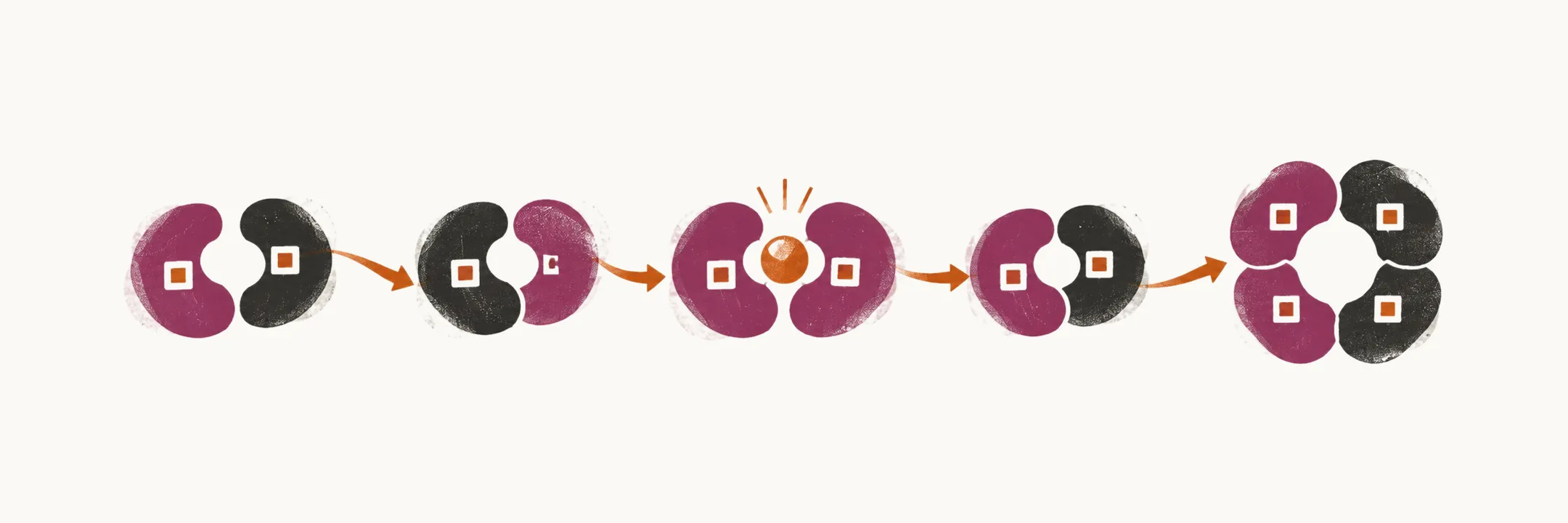
Hemoglobin is the molecule that carries oxygen inside the red cell, and the globin genes that build it are the same genes that are changed in the hemoglobinopathies, the inherited diseases of hemoglobin. Much of what is known about hemoglobin synthesis and function comes from it being unusually easy to study: red cells are accessible for looking at the finished protein, reticulocytes (immature red cells) for following hemoglobin biosynthesis, and leukocytes for checking the mutations that concern hemoglobin.
Red cell production is dynamic in place and in the hemoglobin it makes. It starts in the yolk sac, moves to the fetal liver, and finally settles in the bone marrow, and the type of hemoglobin each cell makes depends on which globin genes are switched on in that cell at that time.
How hemoglobin is built
Hemoglobin is made up of two pairs of dissimilar globin chains, each of which has a tetrapyrrole heme group. The molecule carries oxygen from the lungs and releases it in other tissues.
There are two globins, and for each of them there is one cluster of genes. The clusters are described first, because every chain used in embryonic, fetal and adult life comes from one of them.
The α-globin gene cluster
The α-globin gene cluster is located on the short arm of chromosome 16. Its genes are:
- the embryonic ζ-globin gene, or HBZ
- the duplicated α-globin gene, or HBA1 and HBA2
The β-globin gene cluster
The β-globin gene cluster is located on the short arm of chromosome 11. Its genes are:
- the embryonic ε-globin gene, or HBE
- the two nearly identical fetal γ-globin genes, or HBG1 and HBG2
- the major adult β-globin gene, or HBB
- the minor adult δ-globin gene, or HBD
Fetal and adult hemoglobin
The chains made by these genes combine in different pairs at different ages. Fetal hemoglobin is α2γ2, two α chains with two γ chains, and it is different from the embryonic hemoglobins. Its production is initiated from the 6th week of gestation, reaches its highest fraction around the middle of gestation, and then falls, to less than 1% six months after birth.
Fetal hemoglobin exists to take oxygen from the mother. It binds oxygen more avidly than adult hemoglobin, so at the low oxygen tension of the placenta it can still load oxygen from the maternal blood, and the fetus is supplied across the placenta as a result. The γ chains bind 2,3-bisphosphoglycerate, the molecule inside the red cell that normally lowers hemoglobin’s affinity for oxygen, less well than β chains do, and this is what keeps fetal hemoglobin’s affinity high.
The production of adult hemoglobin, or HbA, which is α2β2, has a reciprocal pattern to the production of fetal hemoglobin: as fetal hemoglobin falls after birth, adult hemoglobin rises.
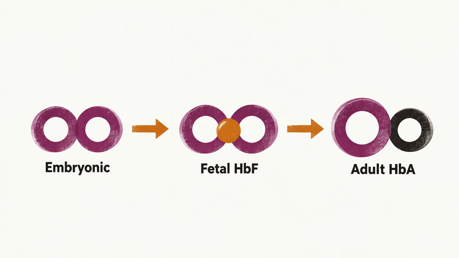
In a normal adult the composition of all the hemoglobin is:
- 96% HbA
- about 1% fetal hemoglobin
- about 2-3% HbA2, which is α2δ2
HbF and HbA2 are functionally not important in the adult, but from a diagnostic point of view they are important: a change in the structure or the amount of one chain alters this normal composition, and that alteration is what the hemoglobinopathies show.