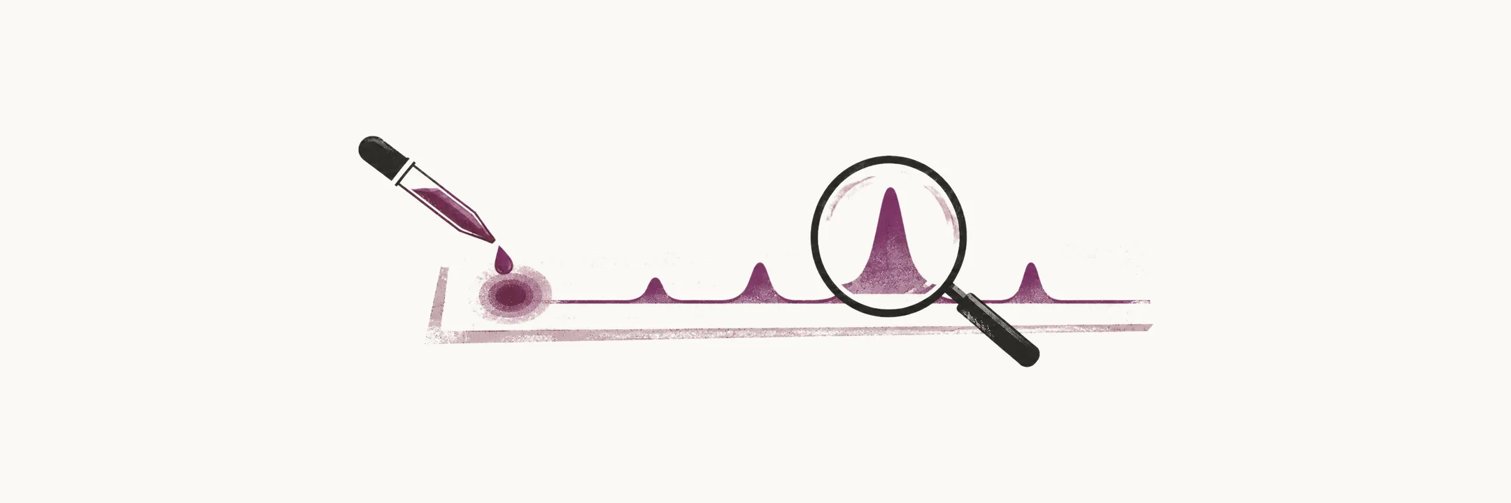
Sickle cell disease (SCD) is diagnosed by looking directly at the hemoglobin a person makes, most often before any symptom appears. The disease is silent at birth: the newborn’s red cells are still full of fetal hemoglobin (HbF), which does not polymerise, that is, does not form the polymers that make red cells sickle, and life-threatening events can begin from about three months of age. Diagnosis therefore depends on screening, and on tests that can separate HbS from the hemoglobins it resembles.
Newborn screening
Newborn screening for hemoglobinopathies is universal in many countries, and in the United States it became standard in all 50 states and the District of Columbia by 2006. A few drops of blood on a filter card are analysed by high-performance liquid chromatography (HPLC) or isoelectric focusing (IEF), the two methods used for the first-line screen. Both separate the hemoglobin fractions in the sample and quantify them, so an abnormal pattern — such as HbS with no HbA — can be recognised even in the first days of life.
The point of finding the disease this early is what can then be prevented. Simple measures started in infancy, such as penicillin prophylaxis, vaccination and education of the parents, avoid most of the early life-threatening complications of sickle cell disease.
Confirmatory testing
A screening result is provisional. The diagnosis is confirmed by a second, different method — hemoglobin electrophoresis or HPLC — and the confirmatory sample is preferably taken before three months of age.
What the confirmatory test shows is the proportion of each hemoglobin, and that proportion is what identifies the genotype:
- in HbSS, HbS is the dominant hemoglobin and HbA is absent;
- in HbSC and in HbS/β-thalassemia, HbS is present together with a second abnormal or reduced fraction;
- in sickle cell trait, HbS makes up roughly 35-45% of the hemoglobin and HbA makes up the majority of the rest.
The last pattern is the one that must not be confused with disease. In sickle cell trait the normal allele still makes HbA, so the HbS fraction stays below the HbA fraction, whereas in HbSS there is no HbA to compete with it.
The solubility test and its limits
The sickle cell solubility test, often called a sickle prep, is the common screening test for HbS in adults, and it is inexpensive, rapid and highly sensitive. It works because HbS is insoluble and precipitates when a reducing agent is added to the sample, so a positive result means HbS is present.
What it cannot do is quantify HbS. A positive solubility test therefore cannot tell sickle cell trait from sickle cell disease, and it needs a confirmatory test to do so. It is also not valid in the newborn, because the high level of fetal hemoglobin dilutes the HbS and produces false negatives.
When the pattern is not enough
Protein-based tests can miss or misclassify several hemoglobinopathies, especially when a β-thalassemia mutation is involved, and a rare variant can migrate like HbS and be mistaken for it. When the fractionation pattern is ambiguous, DNA analysis of the β-globin gene, HBB, is used to establish the genotype.
Prenatal diagnosis uses DNA methods for the same reason, through chorionic villus sampling or amniocentesis. Fetal hemoglobin has no β chain, so the protein itself cannot show whether the β-globin gene is affected before birth; only the gene can.
Once the genotype is established, prevention and treatment can begin: drugs that act on the disease itself, and supportive care such as infection prevention in the patient who has lost splenic function.