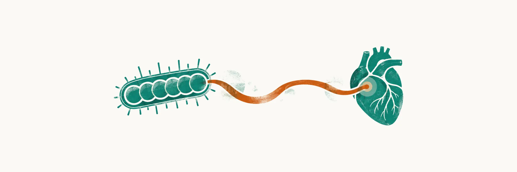
Streptococcus pyogenes, or group A Streptococcus (GAS), is a Gram-positive coccus that grows in chains. It accounts for roughly 90% of bacterial pharyngitis, and its importance is out of proportion to that share because it causes two different kinds of complication, only one of which antibiotics remove. Both kinds make sense once the organism’s equipment is known: what lets it colonise the throat, evade the immune system and spread, and which of its products leave an antibody response behind.
The virulence factors behind the clinical picture
GAS colonises the throat or skin, defends itself against innate and adaptive immunity, and then spreads. The same organism can therefore produce very different illnesses — pharyngitis, scarlet fever, impetigo, cellulitis, necrotising fasciitis, toxic shock, puerperal sepsis — and the variety is easier to hold together as a set of factors, each with a consequence. They fall into three groups.
Surface defences keep the organism from being recognised and engulfed by phagocytes.
- Capsule, made of hyaluronic acid. Because the polymer resembles host connective tissue, it is poorly recognised by the immune system; together with the M protein it gives the organism its antiphagocytic surface.
- M protein, encoded at the emm locus and type-specific. Two polypeptide chains in an α-helix, with a conserved C-terminal end anchored to the cytoplasmic membrane and a variable N-terminal end. It is a protective antigen, but only for its own type: re-infection with the same M type gives type-specific immunity, and another M type does not. Because the N-terminal sequence varies so much between strains, GAS is classified into more than 200 emm types.
- C5a peptidase — degrades C5a, the complement fragment that recruits and activates phagocytes, blunting the local inflammatory response.
Toxins damage host cells or drive the inflammatory response.
- Streptococcal pyrogenic exotoxins (SPE), also called erythrogenic toxins — the best known are SPE-A and SPE-C. Encoded by bacteriophages (viruses that infect bacteria) carried by lysogenic strains, they act as superantigens, bypassing normal antigen presentation and triggering mass cytokine release. They produce the erythema of scarlet fever and contribute to shock and organ failure.
- Streptolysin S — oxygen-stable, serum-soluble; lyses red cells, leucocytes and platelets and stimulates lysosomal enzyme release. It is not immunogenic, so it leaves no antibody trace.
- Streptolysin O — oxygen-labile, cytolytic for leucocytes, platelets and erythrocytes, and immunogenic: antibodies against it are what the antistreptolysin O titre (ASO, also called TAS) measures.
Spreading enzymes break down clots, pus and tissue so that the organism can move through them.
- Streptokinase — two forms that lyse blood clots and contribute to rapid tissue spread of the organism.
- Deoxyribonucleases (DNase A–D) — not cytolytic, but they depolymerise free DNA in pus and so promote spread, and they induce antibody production.
- Hyaluronidase — degrades extracellular matrix hyaluronic acid, again enabling spread.
The serology follows from the immunogenicity differences. Anti-streptolysin O rises after pharyngitis and is the classic marker of a recent streptococcal infection; anti-DNase B and anti-streptokinase are the alternatives and are particularly useful after skin infection, where the ASO response is weaker. Streptolysin S, the tissue-damaging toxin, leaves no antibody at all.
Two kinds of complication, separated by their timing
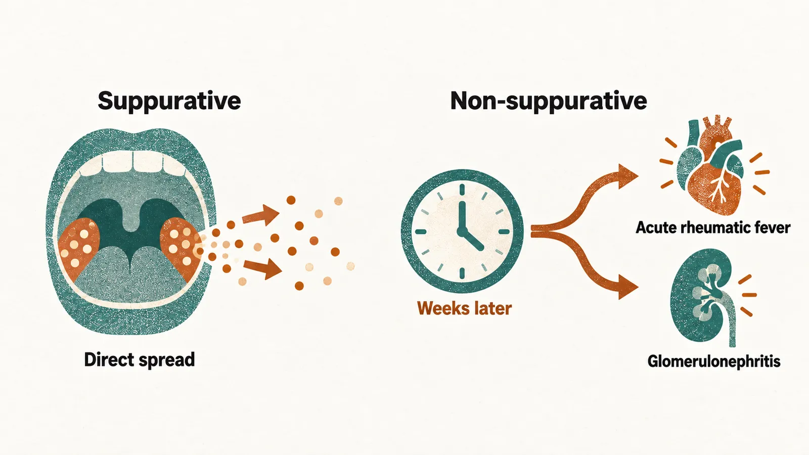
The complications of GAS divide by mechanism. Suppurative (pus-forming) complications come from the organism itself, by direct spread or bloodstream seeding. Non-suppurative complications come from the antibody response to it, and so appear weeks later.
| Suppurative — direct spread or bloodstream seeding | Non-suppurative — immune-mediated, appearing weeks later |
|---|---|
| Peritonsillar cellulitis or abscess, retropharyngeal abscess, cervical lymphadenitis, otitis media, sinusitis, meningitis, brain abscess | Acute rheumatic fever (roughly 1–5 weeks after the sore throat) and acute post-streptococcal glomerulonephritis (roughly 10 days after pharyngitis, up to 3 weeks after a skin infection) |
Both non-suppurative complications rest on molecular mimicry: streptococcal antigens resemble proteins of human heart and kidney, so antibodies raised against the bacterium also recognise the patient’s own tissue. The rheumatic consequence is the reason a sore throat is treated at all in a high-income setting where the disease looks trivial, and it is the reason the ten-day course exists. Scarlet Fever covers the same organism presenting with a rash, and Complications and Pitfalls of Childhood Exanthems follows rheumatic fever and glomerulonephritis in more detail.
Invasive group A streptococcal disease
Most GAS disease is mucosal and self-limiting, but the organism can invade and produce sepsis, necrotising fasciitis or streptococcal toxic shock syndrome, which carries a high mortality; the superantigen exotoxins described above contribute to the shock and organ failure. In 2022 the World Health Organization and the European Centre for Disease Prevention and Control reported an unseasonal increase in invasive GAS disease and scarlet fever in children under 10 years across several European countries, including France, Ireland, the Netherlands, Sweden and the United Kingdom, with a number of deaths. The increase followed a period of unusually low transmission, and the organisms involved have remained susceptible to penicillin. Surveillance of the circulating emm types, the M-protein types described above, continues.
Finding the organism
Virulence factors, antibody responses and emm types are invisible at the bedside, and the signs of GAS pharyngitis overlap with those of adenovirus and Epstein–Barr virus. Establishing that the organism is present is therefore a microbiological question, and a throat swab is what answers it.