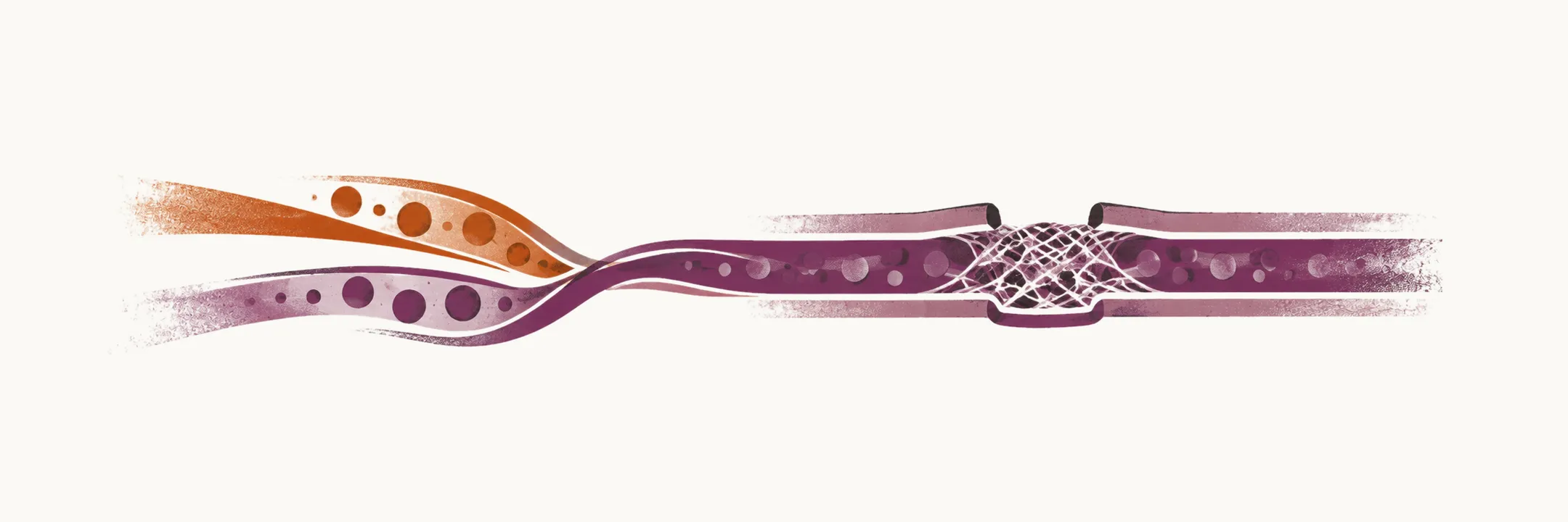
Coagulation is the process that changes liquid blood into a semi-solid clot, and it is made up of two connected stages: primary hemostasis, in which platelets form a plug at the damaged wall, and secondary hemostasis, in which a fibrin mesh stabilises that plug. The stages overlap in time, and the factors activated in each feed back into the other.
Platelet plug formation (primary hemostasis)
The platelet plug is the most rapid reaction to vessel-wall damage, and it is built in four steps.
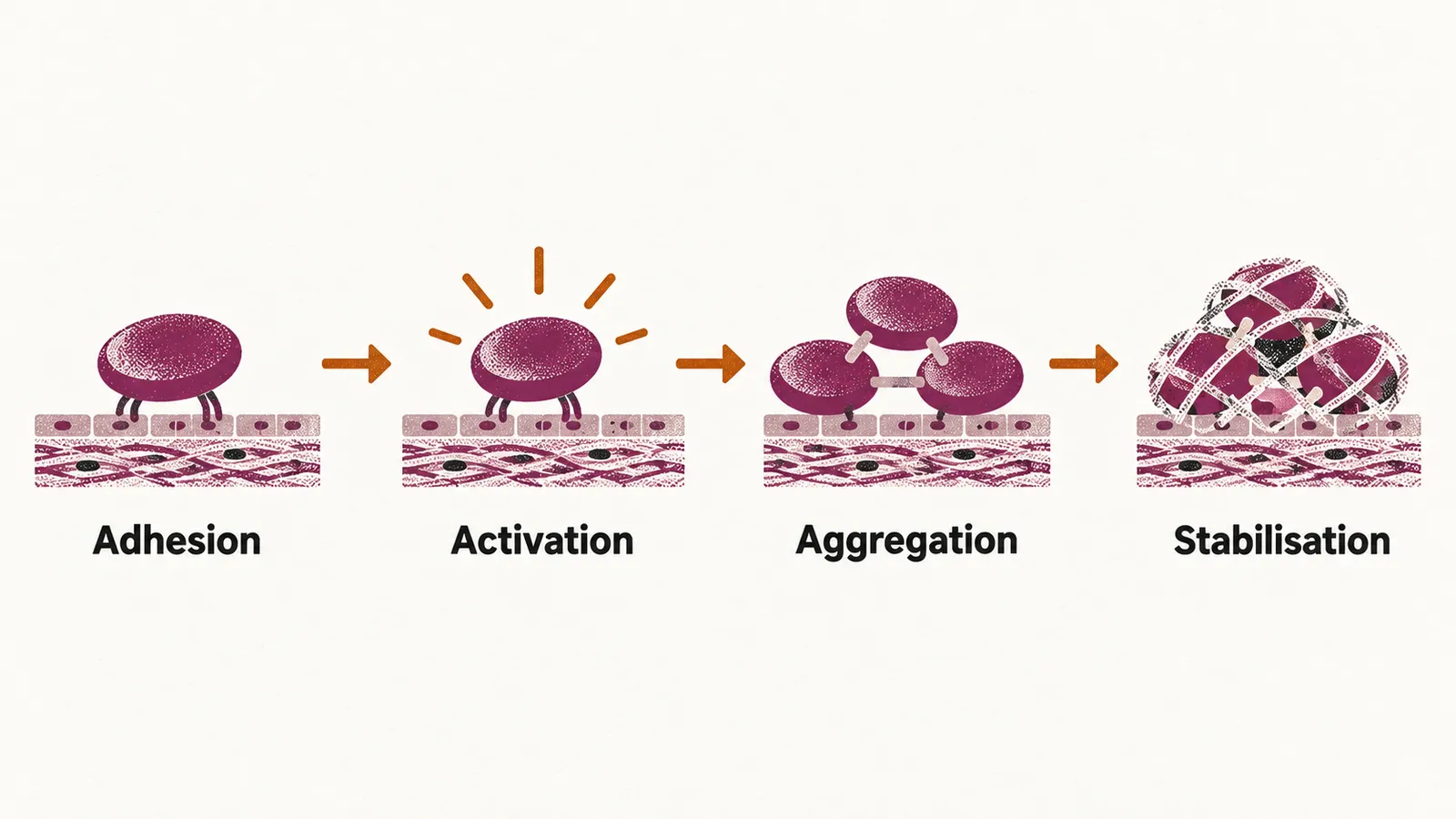
Platelet adhesion. In the normal situation, the shear stress that blood flow produces in the vessels recruits the platelets to the walls and makes them roll along that region; they attach firmly only where mediators that are not normally exposed appear. Adhesion is mediated by attachment of the platelet to:
- von Willebrand factor, or VWF, which is the primary adhesive molecule for the platelet — a kind of molecular glue. It is found in the extracellular matrix of sub-endothelial regions and, in a very small quantity, as a circulating form in the plasma.
- collagen, through the specific platelet receptors for collagen, which attach to the sub-endothelial collagen.
- tissue factor, exposed at the injured surface.
Platelet activation. Two forces switch the platelet on. The first is adhesion itself, which starts activating intracellular signalling pathways inside the platelet as it sticks. The second is humoral mediators: the molecules released by already-activated platelets — ADP, serotonin and calcium — together with epinephrine and thrombin.
ADP acts through its receptors on the platelet, P2Y1 and P2Y12. When they are bound by ADP they activate GPIIb/IIIa and COX-1. GPIIb/IIIa then attaches to fibrinogen, while COX-1 forms TxA2, the major prostaglandin for platelet aggregation.
Platelet aggregation. Once GPIIb/IIIa is active, fibrinogen molecules bridge neighbouring platelets and the plug grows.
Plug stabilisation. The plug is stabilised by forming a fibrin mesh. The most abundant receptor on the surface of the platelet is GPIIb/IIIa, which is normally inactivated but after activation binds fibrinogen and VWF, so it both cross-links platelets and anchors them in the growing plug. The fibrin mesh itself is the product of secondary hemostasis.
Fibrin clot formation (secondary hemostasis)
Coagulation factors, or clotting factors, circulate normally in the inactivated form, as pro-enzymes, so each one has to be switched on by another in a cascade. The cascade is described as an extrinsic and an intrinsic pathway that converge on factor X, but in the body the two run together rather than as separate routes.
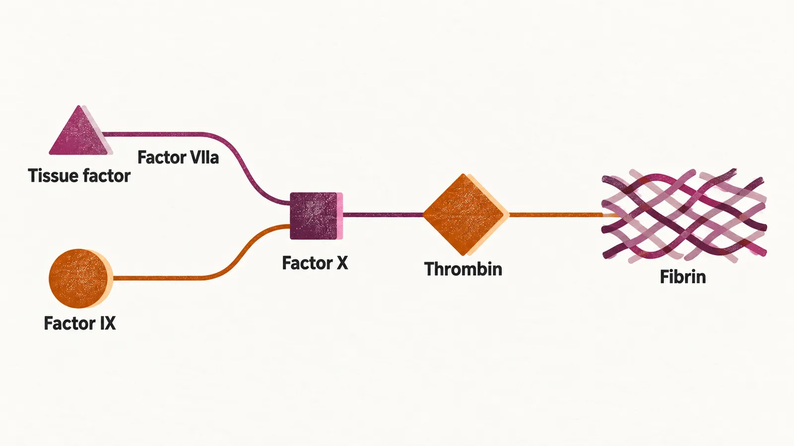
Extrinsic, or tissue factor, pathway. This is the initiator pathway most of the time. Its immediate trigger is the exposure of tissue factor, or TF, which is constitutively expressed in the sub-endothelial cells of the vessels, such as smooth-muscle cells and fibroblasts. TF attaches to the serine protease factor VIIa, and this complex activates factor X.
Intrinsic, or contact, pathway. This is the amplifier pathway that runs after the extrinsic pathway has started, the two working at the same time. Its very first step is the activation of factor XII to XIIa by chemicals that can be found in the blood, high-molecular-weight kininogen (HK) and prekallikrein (PK), although in the normal condition PK and HK are not allowed to start the intrinsic pathway.
The TF–VIIa complex also activates factor X indirectly, by first activating factor IX; factor IXa, with the help of its cofactor factor VIIIa, then activates factor X. Factor IX is activated by factor XI, which is activated by thrombin. Factor XI is more responsible for the propagation of the cascade than for its initiation. Optimal thrombin generation depends on factor IXa, and so on the intrinsic pathway, because the intrinsic pathway can activate factor X much more efficiently than the TF–VIIa complex.
Thrombin. The pivotal protease of the cascade is thrombin, or factor II. It is generated when factor Xa, with factor Va as its cofactor, converts prothrombin. Thrombin is a multifunctional protein, important for:
- activation of the cofactors in the extrinsic pathway, which are factor VIII and factor V;
- activation of factor XIII;
- fibrin formation;
- protein C activation;
- stimulation of the formation of anti-coagulant factors by the endothelial cells.
Both the extrinsic and the intrinsic pathway lead to activation of factor X, which builds the fibrin mesh around the site of vessel-wall damage. Clotting factors do not only activate one another, but also activate the anti-thrombotic factors at the same time, which is the basis of the balance in the hemostatic system.
The fibrin clot forms at the level of the cell membrane of platelets, or even of endothelial cells, specifically at acidic phospholipids. These are not exposed to the outer leaflet of the membrane and therefore have to be transferred from the inner to the outer leaflet.