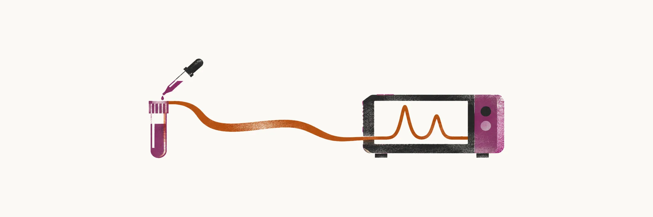
No single laboratory test is able to check all the hemostatic pathways adequately enough to exclude or confirm a bleeding disorder, so a combination of different tests is used instead. The tests are read with the history and examination, and the choice of the next test depends on what the first ones show.
The typical screening panel
A typical screening for a bleeding disorder contains:
- complete blood count (CBC) with platelet count;
- morphology of the platelet;
- screening tests for the coagulation pathways, which are prothrombin time (PT) and activated partial thromboplastin time (aPTT).
These are only the first step of laboratory testing; depending on the result, more and additional tests may be needed. In hematology the results are extremely dependent on pre-analytic variables, such as an incompletely filled tube, the use of a small-gauge needle, a high temperature during sample collection, and a hematocrit over 55%.
Tests for suspected secondary hemostasis disorders
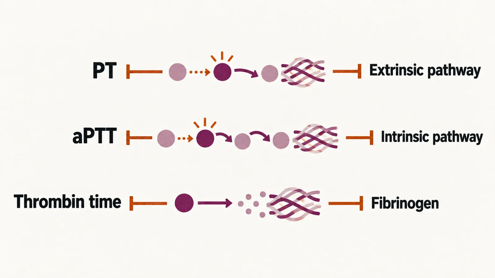
Secondary hemostasis is the fibrin-forming part of clotting. The PT and aPTT screen its two pathways, the mixing study separates a deficiency from an inhibitor, and the thrombin time and reptilase time look at the final step.
Prothrombin time (PT)
Prolongation of the PT indicates extrinsic pathway problems, such as a deficiency or the presence of inhibitors. The substrate that is used to measure the PT is citrated plasma: plasma because it contains the fibrinogen, and citrated so that it is free from the calcium and clotting cannot start until calcium is added back. The PT is therefore the time required for the clotting of citrated plasma after adding calcium and tissue thromboplastin. PT prolongation is seen in the clinics mostly in disseminated intravascular coagulation (DIC), liver diseases, vitamin K deficiency, oral anticoagulant therapy, and deficiency of factor V, VII and X.
For the normalisation of the PT result between different laboratories, which use different tissue thromboplastins, the INR can be used; but the INR is not made for the evaluation of a bleeding disorder per se, it was developed for the monitoring of bleeding due to warfarin and other vitamin K-dependent antagonists.
Activated partial thromboplastin time (aPTT)
Prolongation of the aPTT indicates intrinsic pathway problems, such as a deficiency or the presence of an inhibitor. The substrate is similar to the one used for the PT, which is citrated plasma. The aPTT is the time needed for clotting in that substrate after adding calcium and partial thromboplastin and an activator. Antiphospholipid antibodies, including the lupus anticoagulant, commonly prolong the aPTT and are a leading cause of an unexplained isolated prolongation of the aPTT. In the prolonged aPTT test, the following are considered: DIC; hemophilia A and B; von Willebrand disease (VWD); liver diseases; administration of heparin; massive transfusion of whole blood; and problems related to factor VIII, IX, XI and XII.
Mixing studies
These are tests in which the plasma of the patient is mixed with a specific plasma, which could be completely healthy plasma or plasma that has all the factors except one. They distinguish between a deficiency (in which the missing factor is supplied by the reference plasma and the clotting time corrects) and the presence of an inhibitor (in which the inhibitor also acts on the reference plasma, so the time stays prolonged).
Thrombin time and reptilase time
Both the thrombin time (TT) and the reptilase time (RT) measure the final step of coagulation, which is the formation of the fibrin clot from fibrinogen. They are used in patients who are suspected of fibrinogen deficiency and in patients with prolongation of the aPTT due to the use of an anticoagulant. The TT is sensitive to fibrinogen disorders and also to some drugs, such as heparin, dabigatran and argatroban; the RT is sensitive only to fibrinogen.
Tests for suspected primary hemostasis disorders
If the patient has mucocutaneous bleeding or other reasons to suspect primary hemostasis defects, such as platelet and vascular function defects, the following are also prescribed:
- platelet function analyzer, or PFA-100;
- platelet count and morphology;
- VWF antigen and activity;
- factor VIII activity.
The PFA-100 mimics the blood flow. It has a capillary tube that mimics the blood flow and a reservoir that contains collagen fibres and VWF, beside epinephrine and ADP, so that everything is ready for platelet activation. The end of the tube has a smaller diameter, which leads to a higher shear stress that triggers platelet plug formation, and this activation stops the blood flow; what is measured is the time of plug formation. A prolonged closure time points to a defect in platelet adhesion or aggregation or in VWF.
When the clotting times are normal
If the patient who is suspected of a bleeding disorder has normal clotting times on laboratory tests, the disorders that the screen does not detect have to be suspected: VWD; platelet function abnormalities; vascular and connective tissue disorders; and fibrinolysis disorders.
In the actively bleeding patient
In patients who present with active bleeding, beside the main common laboratory tests related to the bleeding disorder, a fibrinogen activity test is also ordered, because fibrinogen can fall during massive bleeding and replacement depends on the level.