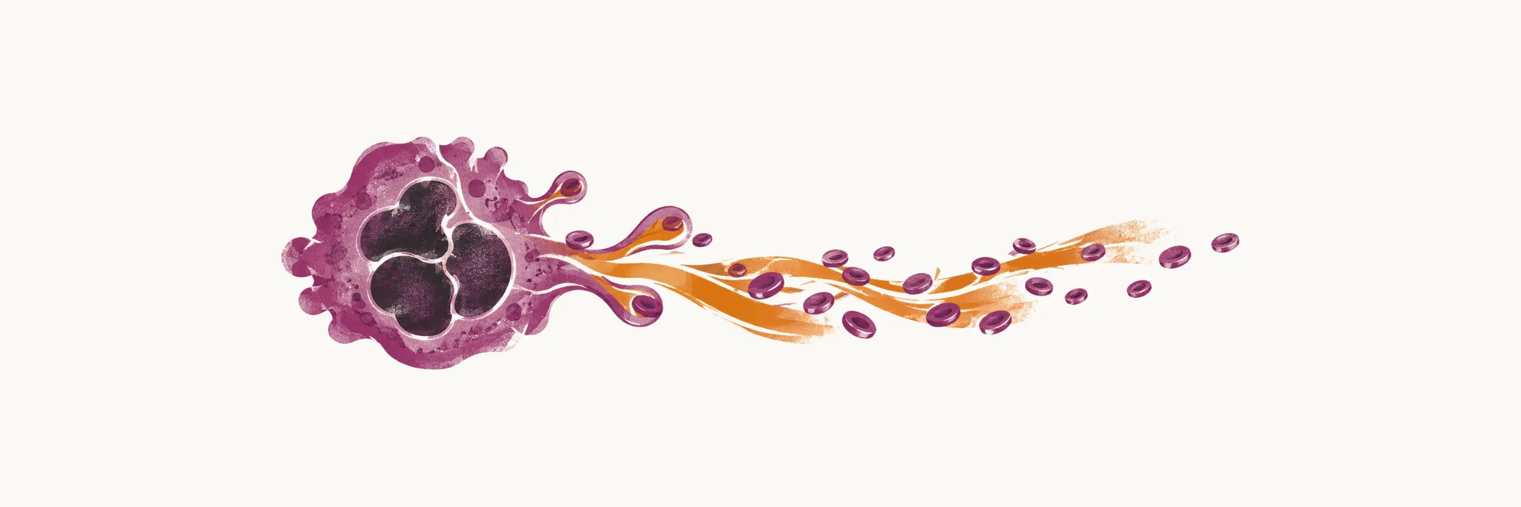
Platelets are the main cellular actor of hemostasis. They are not whole cells but fragments of megakaryocytes, the large precursor cells of the bone marrow, and knowing how they are made explains the counts and measurements used to assess them.
Like all the other blood cells, platelets descend from the hematopoietic stem cells. During platelet biogenesis a sequence of precursor cells is seen, and from the pro-megakaryoblast onwards there is no cytoplasmic division. Instead there is endomitosis, in which the nucleus divides again and again while the cytoplasm never divides, and a huge production of protein. The cell therefore enlarges and accumulates protein without ever splitting into daughter cells, and the cell mass becomes a megakaryocyte.
Platelets in the peripheral blood come from the fragmentation of megakaryocytes during platelet formation in the bone marrow: the cytoplasm of the megakaryocyte is released as platelets.
A few figures are worth remembering because they recur whenever a bleeding disorder is assessed. The size of a platelet corresponds to a mean platelet volume of about 9-11 fL. The normal number of platelets in the peripheral blood is 150-450 × 10⁹ per liter of blood, and their average life is about 7-10 days. The spleen sequesters about 25% of the total platelet mass, which is why splenic enlargement can lower the circulating count.
Platelets do their work at the damaged vessel wall, where they adhere, become activated and form the plug that begins hemostasis.