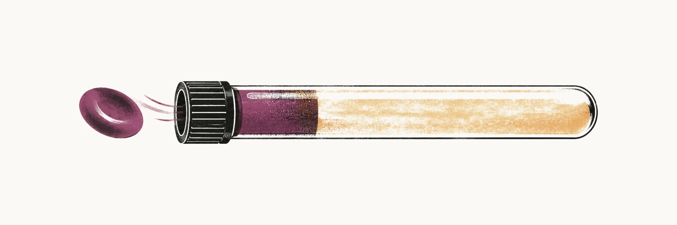
Anemia is defined as a reduced red blood cell mass. That definition is not practical to apply directly to a complete blood count (CBC), so anemia is recognized through one or a combination of the following:
- a reduced hemoglobin concentration
- a reduced red blood cell (RBC) count
- a reduced hematocrit
Clinical consequences and compensation
The clinical consequences of anemia are a decreased delivery of oxygen to the organs and, rarely, hypovolemia, which occurs mostly with acute blood loss.
The body can compensate completely for the anemia, for example by increasing the stroke volume, which masks the signs of anemia. When the compensation is not enough, the clinical picture follows from the tissues that are short of oxygen:
- compensatory signs: dyspnea and tachycardia
- low oxygen to muscle: fatigue, muscle cramps and angina symptoms
- low oxygen to skin and mucosa: pallor, thinning of the hair and nail clubbing
Because compensation can hide the signs, the laboratory numbers are what establish and characterize an anemia.
The indices reported in the CBC
The RBC indices in the CBC describe the red cells and help in the diagnosis of anemia. Three of them, the RBC count (number), the hemoglobin concentration (g/dL) and the hematocrit (%), are the measures by which anemia is recognized. The others describe the individual red cell:
- mean corpuscular volume (MCV, fL): the average volume of the red cells
- mean corpuscular hemoglobin (MCH, pg/cell): the average hemoglobin content per cell; MCH varies largely with the volume of the cell, so the bigger the cell, the more hemoglobin it contains
- mean corpuscular hemoglobin concentration (MCHC, Hb/Ht): the hemoglobin content normalized to volume. MCH gives the content of hemoglobin in each cell, but it does not show whether the amount of hemoglobin inside a given volume of red cells has fallen or risen; the MCHC is the index that answers that question, which makes it more reliable than the MCH for judging the capacity to synthesize hemoglobin
- red cell distribution width (RDW, %)
Classification by MCV
The anemias are grouped by the size of the red cells, which is what the MCV reports:
- microcytic anemia: MCV less than 80 fL
- normocytic anemia: MCV between 80 and 100 fL
- macrocytic anemia: MCV bigger than 100 fL
The three mechanisms of anemia
The MCV group says what the cells look like, and the mechanism says why the red cell mass has fallen. Anemia has three main mechanisms:
- decreased production of RBCs, either a decrease in effective RBC production or ineffective RBC production, in which the cells are made but do not mature
- increased destruction of RBCs, extravascular or intravascular
- blood loss, acute blood loss with hypovolemia or chronic blood loss
The clinical clues that separate these causes are mostly laboratory. For decreased or ineffective RBC production, the reticulocyte count is inappropriately low, meaning low for the degree of the anemia. For increased destruction of RBCs, the reticulocyte count is high, and so are the LDH and the indirect bilirubin, while the haptoglobin is reduced; when the destruction is intravascular, hemoglobinemia and hemoglobinuria are the further clues.
Decreased production is the mechanism of the hypoproliferative anemias, and their commonest cause is iron deficiency, which raises the question of where the body’s iron comes from and how much of it is needed.