
Iron-deficiency anemia is treated with iron, and iron alone is not enough: the loss or the malabsorption that emptied the stores has to be dealt with as well, or the anemia comes back.
Oral iron
The first-line treatment is an oral ferrous salt — ferrous sulfate, ferrous fumarate or ferrous gluconate — starting with one tablet a day. A tablet of ferrous sulfate is 325 mg of the salt and provides 65 mg of elemental iron. If the daily tablet is not tolerated, the options are a reduced dose of one tablet every other day, another oral preparation, or parenteral (intravenous) iron.
Iron is absorbed best on an empty stomach, about 30 minutes before a meal. Food and antacids reduce absorption, and so do tea, coffee, calcium and the phytates of bran and whole grains. Ascorbic acid, either as a 500 mg tablet or as orange juice, enhances absorption without increasing gastric distress.
The adverse effects are gastrointestinal: nausea, epigastric discomfort and constipation, with dark stools. They are the usual reason treatment fails, and larger or more frequent doses do not help: iron raises hepcidin, which blocks the absorption of the next dose, so the extra iron is largely unabsorbed while the side effects increase.
When oral iron is not enough
Parenteral iron is used when oral iron is contraindicated, ineffective or not tolerated. It is considered at an early stage when oral iron is judged unlikely to work, as in malabsorption or in blood loss that continues faster than the gut can replace it, and when the correction of the anemia is urgent; it acts more quickly than oral iron. Transfusion of packed red cells is occasionally required for symptomatic anemia, and iron replacement is still necessary afterwards.
Treating the cause and preventing recurrence
Replacing iron without pursuing the cause is poor practice. In men and postmenopausal women with newly diagnosed iron-deficiency anemia, the gastrointestinal tract is investigated, usually with gastroscopy and colonoscopy, because about a third of them have an underlying pathological abnormality and a gastrointestinal cancer can present this way. Iron replacement is not deferred while the investigations are arranged, unless colonoscopy is imminent.
Where the deficiency is dietary, or where it is likely to recur, the diet can be improved: iron-rich foods, vitamin C taken with them, and tea, coffee, calcium and bran kept away from iron-rich meals. Calcium and iron supplements are taken at different times of the day. Prevention matters most in the groups with the highest iron requirement, such as young children, adolescents, pregnant women and women with heavy menstrual bleeding.
Monitoring the response
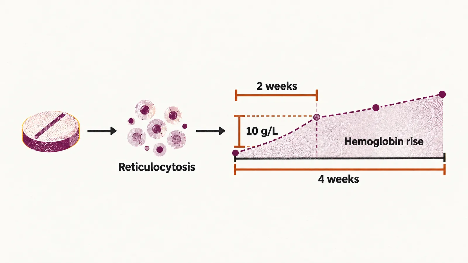
The chronological changes after iron is started are first a reticulocytosis and then an increase in the hemoglobin. The hemoglobin response is monitored over the first 4 weeks, and a rise of at least 10 g/L within 2 weeks is good evidence that the anemia really was due to iron deficiency. The course is continued for about 3 months after the hemoglobin normalizes, and until the serum ferritin comes back to its normal value, so that the marrow iron stores are refilled rather than only the hemoglobin corrected.
When the hemoglobin does not rise as expected, the usual reasons are continuing blood loss, iron that is not being taken or not being absorbed, and a diagnosis other than iron deficiency. That last possibility is why the other causes of a microcytic hypochromic anemia have to be known.