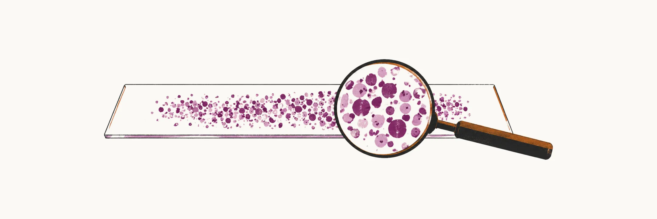
Iron-deficiency anemia is diagnosed from the blood count and the peripheral blood smear in almost every case, even though the bone marrow iron stain remains the gold standard.
The gold standard and what is used instead
The gold standard diagnostic test for iron-deficiency anemia is the amount of iron in the macrophages of a bone marrow biopsy, shown with the Prussian blue stain. It is almost always left undone; instead, the confirmation rests on the CBC and the peripheral blood smear together with the iron indices below.
The indices that confirm iron deficiency
| Index | Normal value | Change in iron deficiency | Caveat |
|---|---|---|---|
| serum iron | 50-150 microgram/dL | decreased | it fluctuates a lot, and it is also decreased in other diseases such as chronic disease, so it is neither a sensitive nor a specific index |
| serum transferrin (TIBC) | TIBC 300-360 microgram/dL | increased, as a compensatory mechanism | TIBC is an indirect way of measuring circulating transferrin |
| transferrin saturation (serum iron/TIBC) | 25-50% | mostly lower than 20% | a saturation over 50% can indicate a disproportionate delivery of iron to non-erythroid tissues rather than to the bone marrow, which can lead to tissue toxicity and iron overload in them |
| serum ferritin | about 100 microgram/L in an adult male and about 30 microgram/L in an adult female, on average, and dependent on sex and age | decreased | the best of the four, highly specific, with a limited sensitivity that is still acceptable; the problem is that it is an acute-phase reactant, so inflammation such as pneumonia raises it |
Serum iron is the circulating iron bound to transferrin, and TIBC measures transferrin indirectly; the ratio of serum iron to TIBC is the transferrin saturation. In iron deficiency there is less iron and more transferrin, so the saturation falls. Ferritin differs from the other three indices because it is a storage protein, so it reflects the iron in the stores.
Hemosiderin and ferritin both store iron in the cells, but ferritin is soluble in the cells while hemosiderin is insoluble and is made mostly when the amount of iron in the tissue exceeds the capacity of its ferritin to store it. Under steady-state conditions the level of serum ferritin correlates with the total store of iron in the body, which is what makes it the most useful index; it depends on sex and age as well.
Red cell protoporphyrin and soluble transferrin receptor
Two further indices add to the picture. Red blood cell protoporphyrin is an intermediate in the production of heme, so its level rises when heme synthesis is impaired, as it is in iron deficiency. Its normal value is less than 30 microgram/dL of red blood cells.
Soluble transferrin receptor is released into the serum from the transferrin receptors on the cells of the bone marrow, so its serum level is indicative of the bone marrow mass; it also rises because the bone marrow cells over-express their receptors to capture more iron from the serum. Its normal value is 4-9 mg/L (4-9 microgram/mL).
The bone marrow iron stores
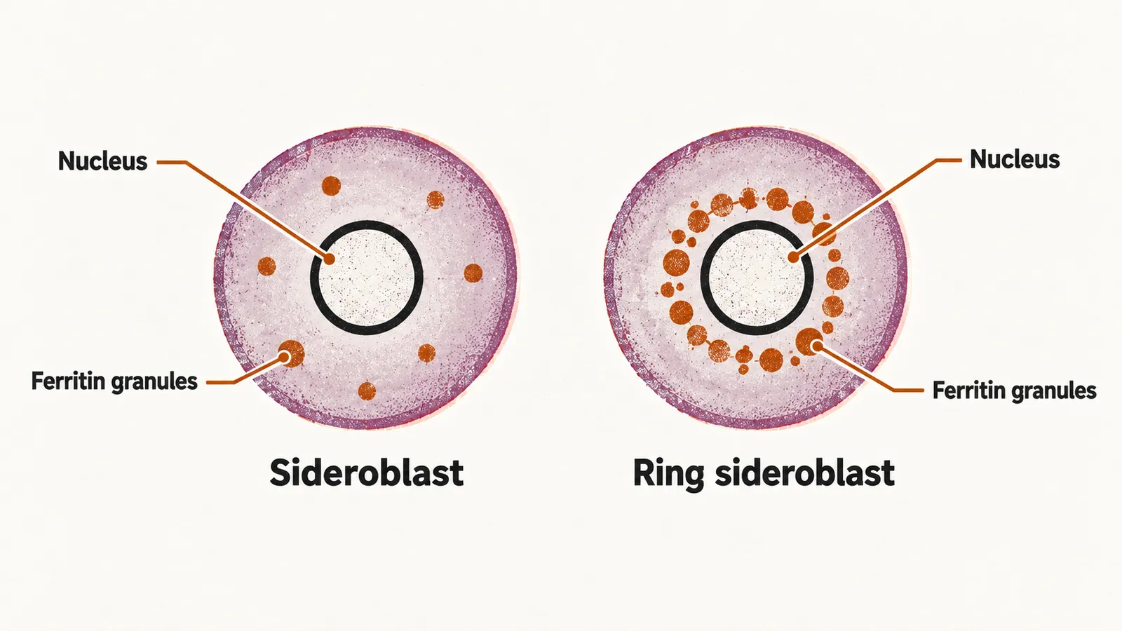
The bone marrow iron stain, done on a bone marrow biopsy, is used mainly to measure iron overload in the body and to monitor the delivery of iron to the erythroblast pool; for measuring the total iron store, the serum ferritin is the better test. In normal conditions, 20-40% of the erythroblasts have visible granules of ferritin, and these cells are called sideroblasts. When the transfer of iron from the RE system to the erythroblasts is blocked, as it is in inflammation, RE iron is seen but few or no sideroblasts are found. In myelodysplastic syndrome a mitochondrial dysfunction can also leave granules of iron arranged around the nucleus, the ring sideroblasts.
Once the diagnosis is confirmed, the question becomes how to replace the iron and how to read the response to that replacement.