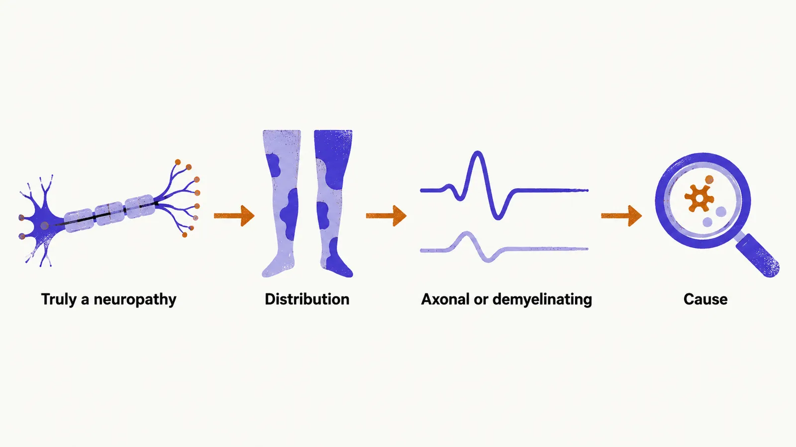
Diagnosing a neuropathy means answering four questions in order: is this truly a neuropathy, what is its distribution, is it axonal or demyelinating, and what is the cause. Skipping ahead to tests before the history and examination have framed these questions is how cases get misdiagnosed.
History
Onset and tempo come first. Weakness evolving over days points to Guillain-Barre syndrome, porphyria, or vasculitis. Subacute progression over weeks to months suggests toxins, paraneoplastic disease, or nutritional deficiency, while chronic courses over months to years fit diabetes, CIDP (chronic inflammatory demyelinating polyradiculoneuropathy), or inherited disease.
Two further clues can settle the question early. Recent respiratory or gastrointestinal infection in the preceding weeks strongly supports Guillain-Barre syndrome. Family history of neurological or even apparently unrelated neuromuscular disease can unmask a mislabeled inherited neuropathy.
The remainder covers exposures and context: chemotherapy, amiodarone, alcohol, solvents, and environmental metals; diabetes, thyroid, renal, liver, HIV, and hepatitis C status; and vegetarian or vegan diets, which raise the risk of B12, B6, and E deficiency.
Examination
The examination establishes distribution, fibre types, and deficit pattern, as taught under foundations. The pattern found points toward particular disorders.
| Finding | Points toward |
|---|---|
| Symmetric stocking-glove sensory loss | Length-dependent polyneuropathy such as diabetes, alcohol, or Charcot-Marie-Tooth disease |
| Asymmetric stepwise nerve involvement | Mononeuritis multiplex: vasculitis, cryoglobulinemia |
| Proximal plus distal weakness | CIDP, Guillain-Barre syndrome, diabetic radiculoplexoneuropathy |
| Pure motor weakness without sensory loss | Multifocal motor neuropathy |
| Prominent autonomic signs | Amyloidosis, diabetes, Guillain-Barre syndrome |
| Pes cavus with hammer toes | Hereditary neuropathy |
Localizing root lesions
Pain and sensory loss in one limb can come from the nerve root (radiculopathy) or from the peripheral nerve itself, and the patterns overlap. A C8 root lesion and an ulnar nerve lesion both affect the medial hand, but the root lesion also involves territories the ulnar nerve never supplies. Mapping the exact sensory boundary, testing muscles outside the suspect nerve, and using needle electromyography (EMG) to place the lesion resolve the question.
When root disease competes with nerve disease, three lumbar roots cover most questions.
| Root | Sensory territory | Key motor deficit | Reflex |
|---|---|---|---|
| L4 | Anterior thigh, medial leg and foot | Quadriceps weakness | Reduced knee jerk |
| L5 | Lateral leg, foot dorsum, great toe | Foot drop with steppage gait | No specific reflex |
| S1 | Posterior leg, lateral ankle, sole | Weak plantar flexion | Reduced or absent ankle jerk |
Root pain often starts proximally in the back or buttock, while mononeuropathy stays focal. The main mimic pairs are L5 radiculopathy with fibular-head peroneal palsy and S1 radiculopathy with tibial disease, resolved by exact sensory mapping, out-of-territory muscle testing, and EMG.
Laboratory testing
First-line testing screens every neuropathy:
- Blood count and inflammatory markers, fasting glucose with HbA1c, and liver, kidney, and thyroid function.
- Vitamins B12, B6, and E.
- Serum protein electrophoresis with immunofixation for monoclonal proteins.
- Antinuclear and related autoantibodies, hepatitis B and C serology, and rheumatoid factor.
Second-line testing follows specific suspicions: anti-GM1 and anti-MAG antibodies for multifocal motor and anti-MAG neuropathies, anti-ganglioside antibodies including GQ1b for Guillain-Barre variants, VEGF for POEMS syndrome, gene panels for inherited disease, and urine testing for Bence Jones protein or systemic amyloidosis.
Nerve conduction studies and EMG
Electroneurography with needle EMG is the central tool for separating axonal from demyelinating disease and for localizing lesions. What the studies measure, how the axonal and demyelinating signatures differ, which thresholds support demyelination, and when the studies are normal are covered under nerve conduction studies and EMG.
Spinal fluid, imaging, ultrasound, and biopsy
Spinal fluid
Spinal fluid matters most for inflammatory disease: Guillain-Barre syndrome and CIDP show raised protein with a normal cell count, though protein can be normal in the first days of GBS. Fluid also screens for infection and inflammation. Details and timing caveats sit under Guillain-Barre syndrome.
Imaging and ultrasound
MRI visualizes root enlargement in CIDP or amyloidosis, cord compression in suspected radiculopathy, and muscle denervation patterns. High-resolution nerve ultrasound detects enlargement at entrapment sites and in inflammatory disease and increasingly complements conduction studies.
Nerve biopsy
Nerve biopsy, usually of the sural nerve, is reserved for situations where it changes management: suspected vasculitis confined to nerves, amyloidosis, sarcoidosis, tumor or lymphoma infiltration, leprosy, and severe unexplained disease. It leaves permanent numbness in the biopsied territory, so it is never a routine step.
The diagnostic flow
The workup runs as a sequence of decisions: confirm peripheral nerve involvement, classify the distribution as symmetric, asymmetric stepwise, or focal, identify the fibre types, establish axonal versus demyelinating mechanism by conduction studies, and then let the history, extranervous examination, and targeted laboratories name the cause. Treatment by cause follows under treatment, and the drugs used for pain under neuropathic pain.