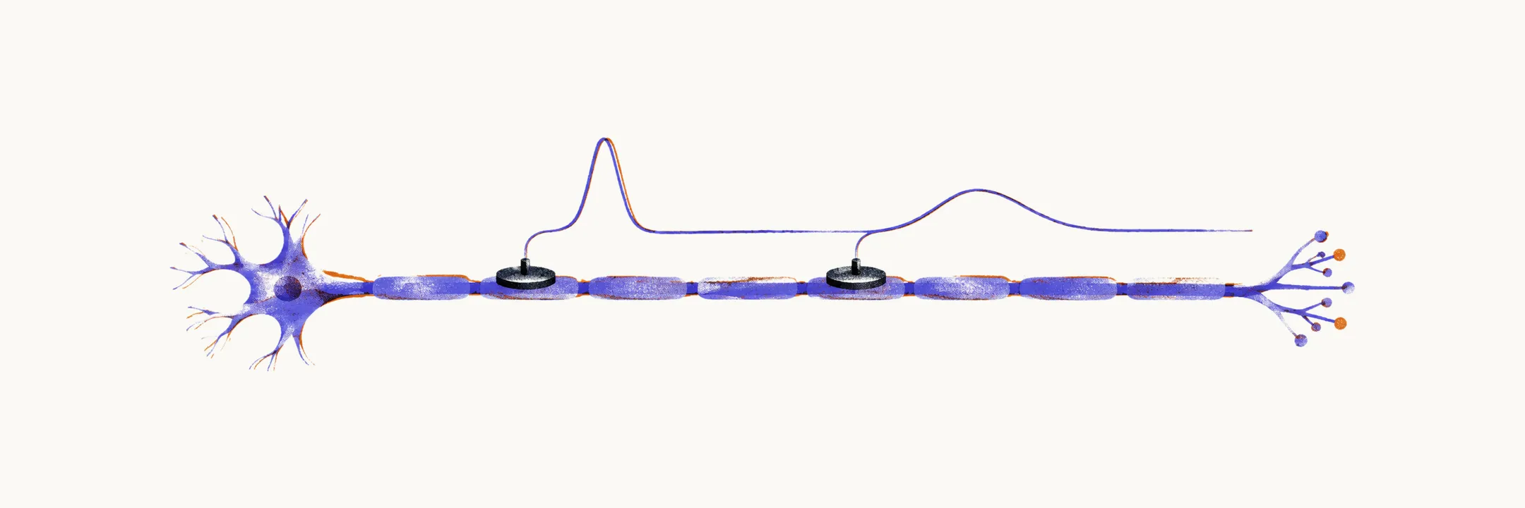
Nerve conduction studies and needle electromyography (EMG) are the central investigations in peripheral neuropathy. Together they answer three questions the clinical examination can only suggest: whether the problem lies in the peripheral nerve, whether the mechanism is axonal or demyelinating, and where along the nerve the lesion sits. They do not identify the cause, which comes from the history, examination, and laboratory testing.
What the studies measure
A motor nerve conduction study stimulates the nerve at two or more points and records the compound muscle action potential from a muscle it supplies. A sensory study records the sensory nerve action potential from the nerve itself. Amplitude reflects how many functioning axons or muscle fibres contribute. Conduction velocity is the distance between stimulation points divided by the latency difference between them, and it is normally above about 50 m per second in the arms. Distal latency is the time from stimulation to the recorded response over the most distal segment, and F-wave latency tests the proximal segment and the nerve root.
The axonal and demyelinating signatures
The two mechanisms leave different fingerprints, and this distinction shapes both interpretation and expected recovery.
| Mechanism | Amplitude | Conduction velocity and latency | Typical diseases |
|---|---|---|---|
| Axonal loss | reduced | relatively preserved | diabetes, alcohol, toxins, Charcot-Marie-Tooth type 2 |
| Demyelination | relatively preserved | slowed velocity, prolonged latencies | Guillain-Barre syndrome, CIDP (chronic inflammatory demyelinating polyradiculoneuropathy), Charcot-Marie-Tooth type 1, anti-MAG neuropathy |
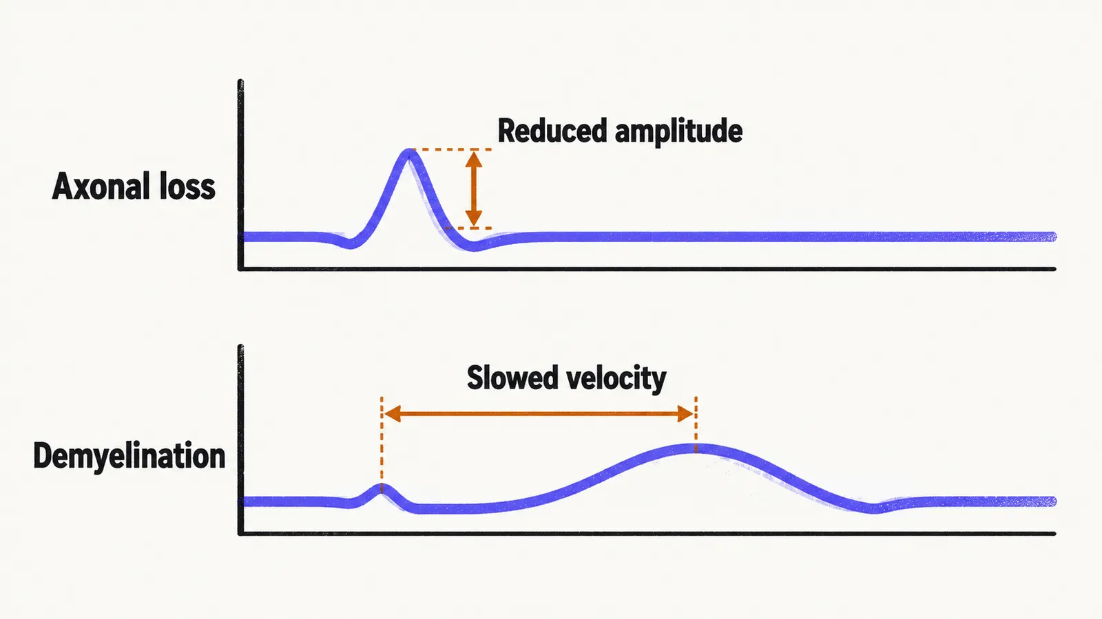
Two additional demyelinating features matter. Conduction block is a fall in the compound muscle action potential amplitude when the nerve is stimulated proximal to a lesion compared with distal to it, which means the signal is being stopped rather than merely slowed. Temporal dispersion is widening and fragmentation of the response as faster and slower fibres desynchronize. Many chronic neuropathies show a mixture of both signatures.
Published criteria put numbers on this. In the current European Academy of Neurology and Peripheral Nerve Society criteria for CIDP, demyelination is supported by a distal motor latency at least 50% above the upper limit of normal, a motor conduction velocity at least 30% below the lower limit of normal, an F-wave latency at least 20% above the upper limit of normal, or a conduction block of at least 30% amplitude reduction, and a definition generally requires such findings in more than one nerve, with the median nerve at the wrist excluded because carpal tunnel syndrome is so common.
Needle EMG
Needle EMG examines the muscle rather than the nerve. Fibrillation potentials and positive sharp waves are signs of active denervation (loss of nerve supply to muscle fibres) and appear 2 to 3 weeks after axonal injury, so an early study can look normal and a repeat study later shows the damage. Fasciculation potentials mark motor unit (a motor neuron with the muscle fibres it supplies) irritability and occur in peripheral neuropathy as well as in motor neuron disease. Changes in motor unit action potentials and reduced recruitment appear as reinnervation proceeds, and paraspinal muscle sampling can show denervation earlier than limb muscles.
What the studies localize
Because conduction can be measured at chosen points along a nerve, the studies place a lesion precisely. Focal slowing across a tunnel identifies an entrapment, such as across the carpal tunnel or behind the medial epicondyle, and it also grades severity, which is why it supports decisions about surgery in carpal tunnel syndrome. Slowing in a single nerve with normal neighbouring nerves suggests a focal process, whereas widespread abnormalities suggest a generalized one.
They also help distinguish root disease from nerve disease. In radiculopathy the lesion lies at or proximal to the sensory root, so the sensory nerve action potential from that nerve is often preserved while needle EMG shows denervation in muscles of the corresponding myotome (the muscles supplied by one root). A lesion of the peripheral nerve itself reduces the sensory response as well. Comparing sensory and motor responses in the same territory is also how the studies separate sensory from motor fibre involvement.
When the studies are normal
A normal study does not exclude neuropathy, and three situations come up repeatedly:
- Small-fibre neuropathy involves fibres below the resolution of the studies, as described under small-fibre neuropathy.
- In Guillain-Barre syndrome the studies are often normal in the first days and become diagnostic in most patients by the second week.
- Mild carpal tunnel syndrome can produce symptoms before conduction slows enough to be recorded.
Limits and complements
Results depend on technique and physiology. Nerve temperature changes conduction velocity, so cool limbs conduct slowly, and age, height, and limb length shift normal values, which is why each laboratory uses its own reference ranges. Amplitude depends on electrode placement and the distance between stimulation and recording sites. Nerve ultrasound complements the studies by showing segmental enlargement at entrapment sites and in inflammatory disease, and it is discussed with imaging under diagnosis.
The studies stop at mechanism and site, so naming the cause, and then acting on it, depends on the history, laboratory testing, and cause-directed treatment.