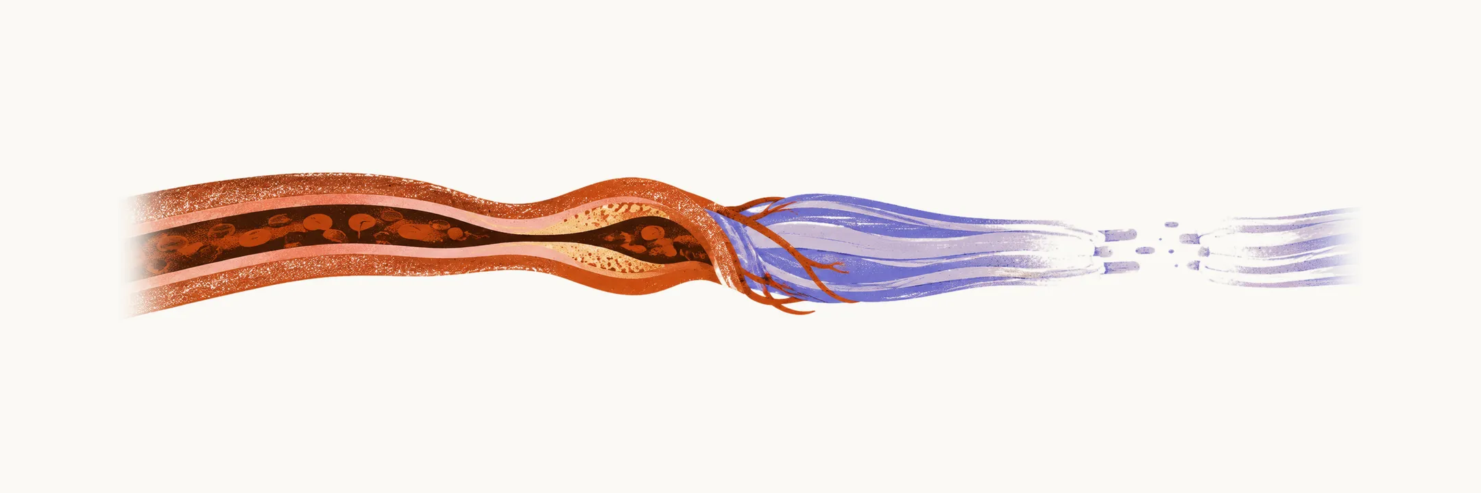
Mononeuritis multiplex means damage to several separate named nerve trunks, appearing at different sites and different times rather than in a length-dependent sequence. A patient might develop a wrist drop, then weeks later a foot drop on the other side. That asymmetric, stepwise course signals a process attacking nerves one by one, and it carries a different differential and urgency from a single entrapment.
Why separate nerves fail
The usual mechanism is ischemia. Small arteries and arterioles supplying a nerve, the vasa nervorum, become inflamed and narrowed, so the nerve trunk infarcts in its own territory. The injury is axonal and starts abruptly, which is why mononeuritis multiplex is typically painful and why weakness or numbness can appear suddenly in one nerve distribution after another. Because nerve trunks rather than nerve endings are affected, the deficits follow the territory of the named nerve rather than the stocking-glove gradient of a polyneuropathy.
Causes
Vasculitis, inflammation of blood vessel walls, tops the list. It may be part of a systemic disease, such as polyarteritis nodosa, granulomatosis with polyangiitis, microscopic polyangiitis, or eosinophilic granulomatosis with polyangiitis, or it may be confined to the nerves, which is called nonsystemic vasculitic neuropathy. Secondary forms accompany rheumatoid arthritis and other connective tissue diseases, Sjogren syndrome, systemic lupus erythematosus, cryoglobulinemia (often hepatitis C related), hepatitis B, and HIV.
Other causes of the same pattern include diabetes, leprosy, sarcoidosis, tumor or lymphoma infiltration, and multifocal immune neuropathies such as Lewis-Sumner syndrome.
Two conditions mimic the pattern without vasculitic infarction. Hereditary neuropathy with liability to pressure palsies is the inherited mimic: ordinary minor pressure produces recurrent focal palsies rather than infarcted nerves, and it does not respond to immunotherapy, as described under hereditary neuropathies. Multifocal motor neuropathy produces a stepwise pattern too, but it is predominantly motor with minimal or no sensory loss, and it is demyelinating rather than axonal, as covered under inflammatory and immune neuropathies.
Clinical recognition
Deficits are asymmetric and sensorimotor in most cases, and pain often precedes or accompanies the weakness. Signs of systemic inflammation, such as fever, weight loss, rash, arthralgia, hypertension, or impaired renal function, belong to the same picture because the underlying vasculitis is often multisystem.
Two distinctions matter most. Entrapment neuropathies are also focal, but they occur at recognized anatomical tunnels and the deficit stays in one nerve; mononeuritis multiplex moves across territories and times. And if the asymmetric deficits gradually merge into a symmetric stocking-glove pattern, the process has become a confluent polyneuropathy, and classification shifts to the framework under polyneuropathy classification.
Investigation
Electrodiagnostic studies show an asymmetric, multifocal, axonal neuropathy, and they identify which nerve is most affected and therefore most useful to biopsy. Nerve biopsy is reserved for situations where it changes management, above all suspected vasculitis confined to nerves. Consensus guidance favors sampling a clinically and electrically affected sensory nerve, and combining a sensory nerve biopsy with an adjacent muscle improves the yield over nerve alone. Definite vasculitic neuropathy requires inflammation within the vessel wall together with vessel wall damage, and because the lesions are patchy, a negative biopsy does not exclude vasculitis.
The systemic search runs alongside: inflammatory markers, antineutrophil cytoplasmic antibodies, cryoglobulins, hepatitis B and C serology, HIV testing, antinuclear antibodies, urinalysis and renal function, and targeted imaging or malignancy workup. Laboratory testing in the general neuropathy workup is described under diagnosis.
Treatment and outcome
Treatment is urgent because the injury is infarction and the aim is to prevent the next nerve from being lost. Corticosteroids are the foundation, and severe, rapidly progressive, or systemic disease adds cyclophosphamide, or rituximab for antineutrophil cytoplasmic antibody-associated vasculitis. Nonsystemic vasculitic neuropathy is treated with steroids first line for at least 6 months, with azathioprine or methotrexate maintenance after combination induction.
Recovery is slow and often incomplete, because regeneration of an infarcted axon proceeds at about 1 mm per day and the longest nerves recover least. Pain control, physiotherapy, and orthotics such as an ankle-foot orthosis for foot drop carry the day-to-day management while immunosuppression takes effect.
Most neuropathies do not follow this asymmetric, nerve-by-nerve course. Symmetric, length-dependent polyneuropathy is the largest group, and it is sorted by a different framework.