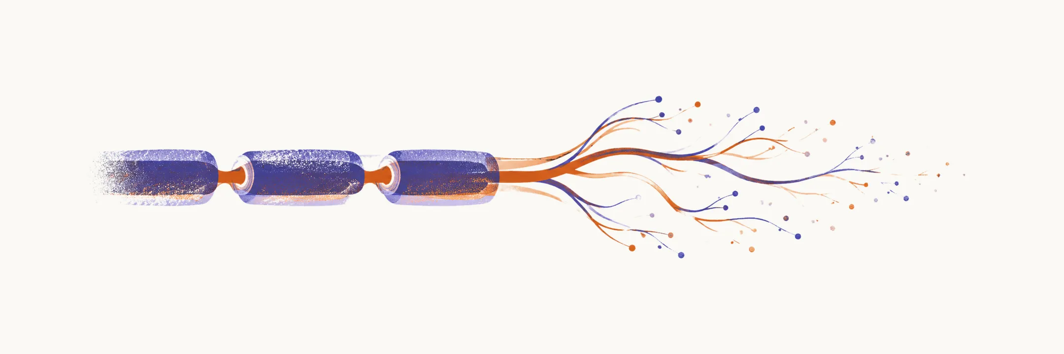
Small-fibre neuropathy damages the thinly myelinated A-delta fibres and unmyelinated C fibres that carry pain and temperature, along with the small autonomic fibres that control sweating and blood pressure. The examination findings that normally confirm a neuropathy can all be normal, because these fibres are not what most tests measure, so the diagnosis rests on the symptom pattern and on a test that routine workups do not include.
Clinical pattern
Pain and altered sensation dominate. Burning feet are the classic complaint, often with stabbing or electric-shock sensations, tingling, and pain that is worse at night. The distribution is length-dependent at first, starting in the feet and rising in a stocking pattern, although non-length-dependent and patchy presentations occur. Allodynia (pain from a normally painless stimulus) and hyperalgesia (exaggerated pain from a painful stimulus) are common, so bedsheets or socks become intolerable.
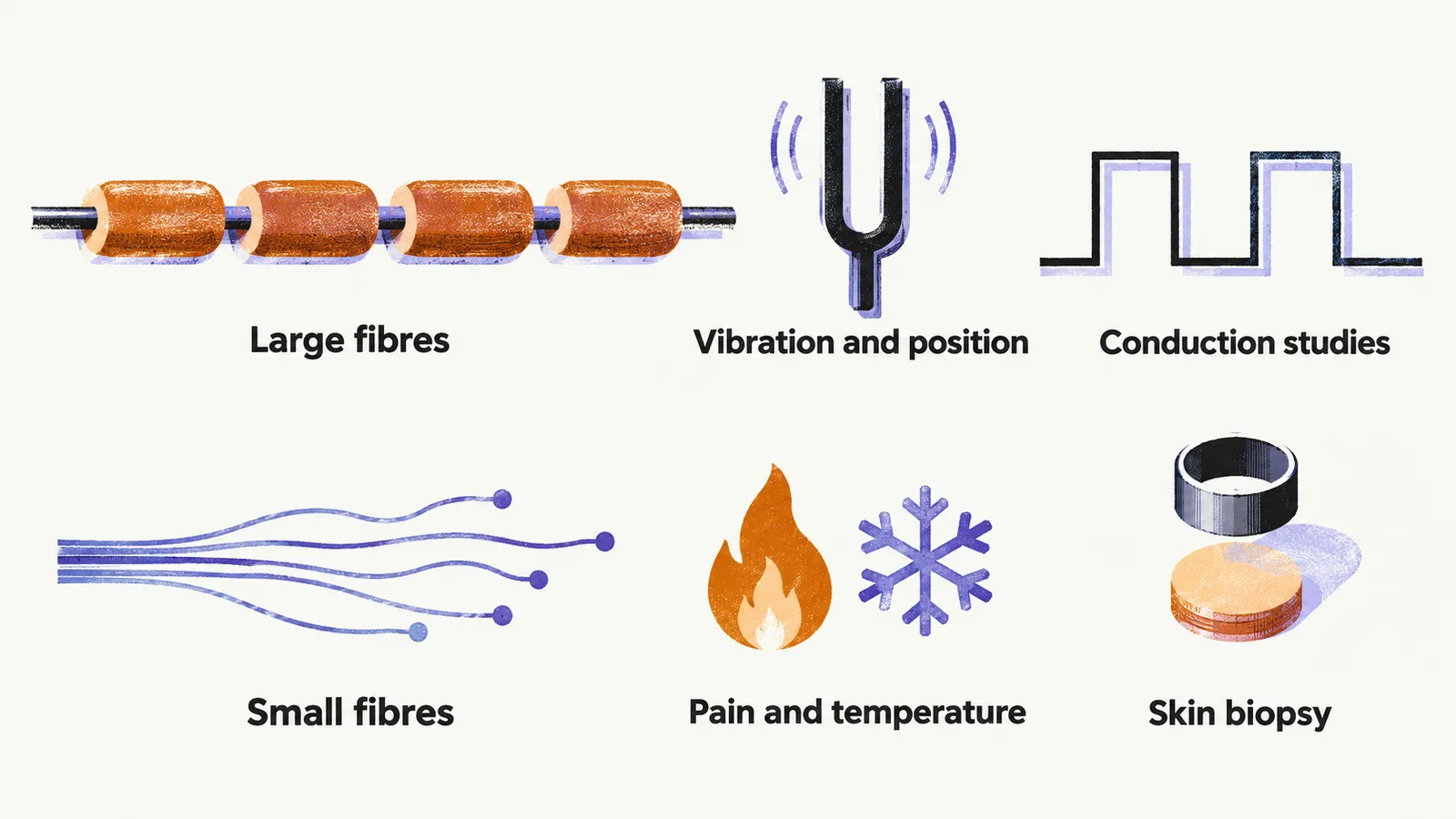
Pinprick and temperature sensation are reduced, and patients often describe an area of numbness they had not reported. Vibration sense, position sense, muscle power, and deep tendon reflexes are typically preserved. Autonomic involvement, when present, brings reduced sweating in the affected area, orthostatic intolerance, gastroparesis, or erectile dysfunction, and it is a signal that the disease is no longer purely sensory.
Why conduction studies are normal
Standard nerve conduction studies and needle electromyography (EMG) examine large myelinated fibres. The thinly myelinated and unmyelinated fibres that carry pain and temperature are below their resolution, so a normal electrodiagnostic study does not exclude small-fibre neuropathy. Quantitative sensory testing addresses them directly by measuring warm and cold detection thresholds, and the skin biopsy described below examines their endings in the epidermis.
Confirmation
The supporting test is a skin biopsy taken with a 3-mm punch at the distal leg, 10 cm above the lateral malleolus, immunostained for intraepidermal nerve fibre density. A reduced density compared with age-matched normative values supports the diagnosis, and consensus guidance gives distal-leg skin biopsy a strong recommendation for length-dependent small-fibre neuropathy. A biopsy from the proximal thigh helps separate length-dependent from non-length-dependent disease, in which the proximal site is also affected.
The tests are not absolute. Reported sensitivity for the biopsy is around three-quarters, with specificity around two-thirds to four-fifths, so a normal biopsy does not rule out the disease, especially in patchy, early, or predominantly autonomic presentations. The biopsy confirms structural loss but rarely identifies the cause, so etiologic testing continues regardless of the result.
Causes
Diabetes is the most common cause, and impaired glucose tolerance and metabolic syndrome are recognized contributors. A substantial proportion of cases remain idiopathic even after thorough testing.
Other causes to consider include alcohol, vitamin B12 deficiency, chemotherapy, HIV and hepatitis C infection, monoclonal gammopathy, hypothyroidism, Sjogren syndrome, celiac disease, sarcoidosis, and drugs such as metronidazole and nitrofurantoin.
Inherited forms exist and are suggested by early onset, a family history, or an unusual distribution: sodium channel variants affecting SCN9A and SCN10A, Fabry disease, transthyretin amyloidosis, and hereditary sensory and autonomic neuropathies.
Investigation and management
Testing is directed at the causes above and follows the same logic as the general neuropathy workup under diagnosis: fasting glucose and HbA1c, sometimes with an oral glucose tolerance test, vitamin B12 with methylmalonic acid where the level is borderline, serum protein electrophoresis with immunofixation, HIV and hepatitis C serology, thyroid and renal function, and autoimmune testing guided by the history.
Management has three strands:
- The cause is treated where one is found, which for diabetes means glycemic and vascular risk control.
- Pain is treated with the agents for neuropathic pain described under neuropathic pain, which act on the symptoms rather than on the disease.
- Because pain sensation is impaired, foot protection matters as much as analgesia, since burns, blisters, and ulcers can go unnoticed.
Some causes, such as diabetes and amyloidosis, later progress to a mixed small- and large-fibre neuropathy.