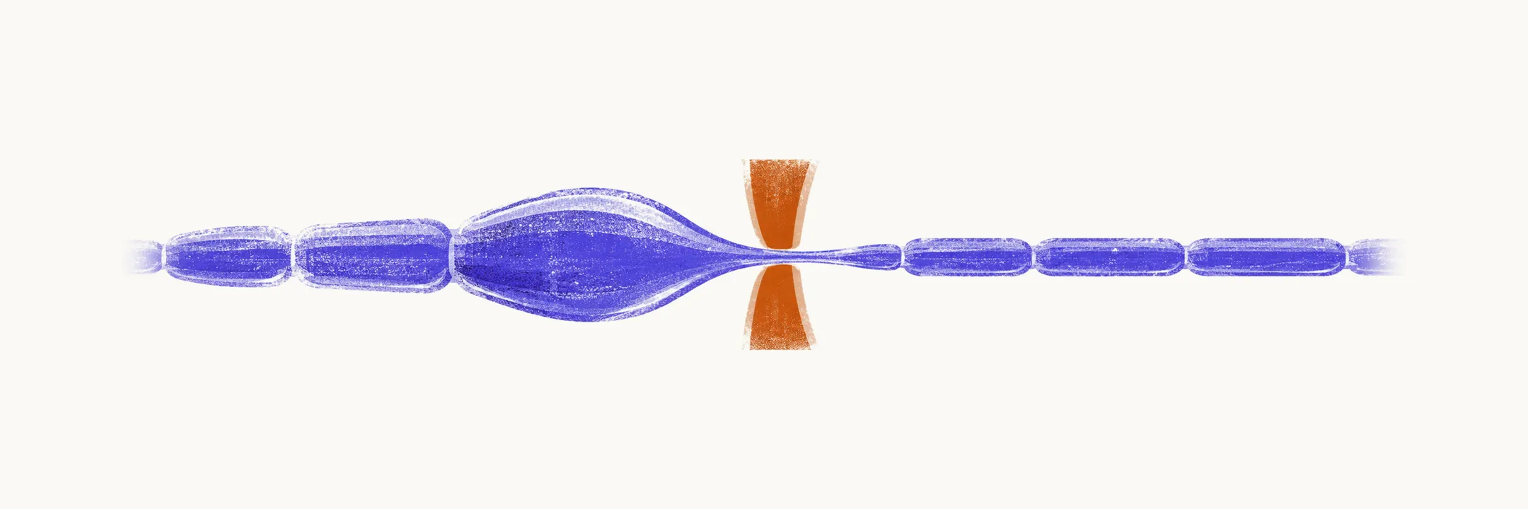
Entrapment neuropathies come from chronic compression of a nerve inside a narrow anatomical tunnel with unyielding walls. Each tunnel produces a characteristic territory of numbness and weakness, so these syndromes test whether the lesion can be localized from symptoms alone.
Carpal tunnel syndrome
The median nerve is compressed beneath the flexor retinaculum, the fibrous band that forms the roof of the tunnel at the wrist. It is the most common focal neuropathy of the upper limb and is more frequent in women, with fluid retention, pregnancy, diabetes, and repetitive wrist loading among the recognized associations.
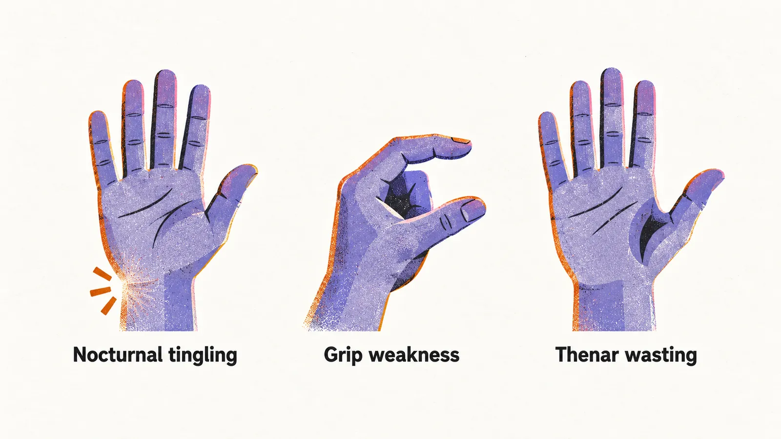
The course typically passes through three phases. First comes nocturnal tingling and numbness in the thumb, index, and middle fingers, with patients waking to shake the hand. Then pain intensifies, grip weakens, thumb opposition falters, and the thenar eminence (the muscle bulge at the base of the thumb) begins to flatten. In advanced disease the thumb muscles waste markedly and the patient cannot oppose thumb to index finger. Pain sometimes eases at this stage, not because the nerve recovered but because severely damaged fibres stop generating ectopic signals.
Three bedside tests support the diagnosis. Direct pressure over the tunnel reproduces tingling, tapping over the median nerve at the wrist sends an electric sensation into median territory (Tinel sign, which marks nerve irritability rather than this specific tunnel), and sustained full wrist flexion for about a minute reproduces numbness (Phalen test). Confirmation and severity grading come from nerve conduction studies, described under nerve conduction studies and EMG.
Treatment starts conservatively with night splinting and, in suitable patients, corticosteroid injection into the tunnel. Surgical division of the retinaculum is the definitive treatment when conservative measures fail or when there is progressive thenar wasting or persistent sensory loss. In practice this means trying conservative care first, without delaying surgery once deficits progress.
Cubital tunnel syndrome
The ulnar nerve is compressed at the elbow behind the medial epicondyle. It is the second most common upper-limb focal neuropathy and considerably less frequent than carpal tunnel syndrome.
Early symptoms are tingling in the little finger and the ulnar half of the ring finger, worse with sustained elbow flexion, which narrows the tunnel. Later comes persistent sensory loss with wasting of the hypothenar eminence (the muscle bulge on the little-finger side of the palm) and the first dorsal interosseous muscle. A positive Froment sign reveals the weakness: asked to pinch paper between thumb and index finger, the patient flexes the thumb IP joint using median-innervated flexor pollicis longus because the ulnar-innervated adductor pollicis is too weak.
Management begins with activity modification and avoiding prolonged flexion, with splinting if symptoms persist. About half of mild cases settle without intervention. Surgical anterior transposition of the nerve is reserved for refractory or progressive cases.
Other single-nerve lesions
The same mechanism of compression at a recognized site produces other single-nerve syndromes, each with its own hallmark.
| Nerve | Syndrome | Hallmark |
|---|---|---|
| Radial nerve at the upper arm | Saturday night palsy | Wrist drop with inability to extend wrist and fingers after prolonged pressure |
| Median nerve in the forearm | Pronator teres syndrome | Forearm ache with weak grip, without the nocturnal pattern of carpal tunnel disease |
| Ulnar nerve at the wrist | Guyon canal syndrome | Hand weakness and sensory loss confined to the hand, sparing the forearm |
| Common fibular nerve at the fibular head | Peroneal compression | Foot drop with steppage gait and sensory loss over the lateral leg and foot dorsum |
| Lateral femoral cutaneous nerve at the inguinal ligament | Meralgia paresthetica | Burning and tingling over the outer thigh, common with obesity and pregnancy |
| Tibial nerve at the ankle | Tarsal tunnel syndrome | Plantar burning pain and numbness, sometimes with a Tinel sign behind the medial malleolus |
Femoral involvement with acute painful thigh weakness in diabetes behaves differently from a simple entrapment and is covered under diabetic neuropathy.
When the pattern is not entrapment
When individual nerves fail one after another at different sites and times, the pattern is mononeuritis multiplex rather than entrapment. A patient might develop a wrist drop, then weeks later a foot drop on the other side. That asymmetric, stepwise course signals a systemic process attacking nerves one by one, and it is covered under mononeuritis multiplex. Recurrent focal palsies after trivial pressure instead suggest the inherited mimic, hereditary neuropathy with liability to pressure palsies, described under hereditary neuropathies.