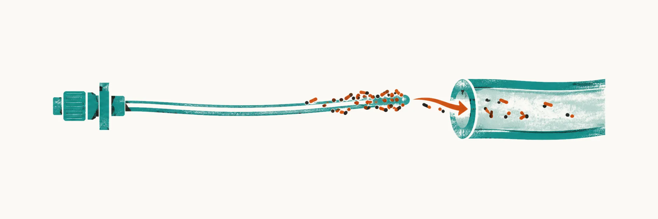
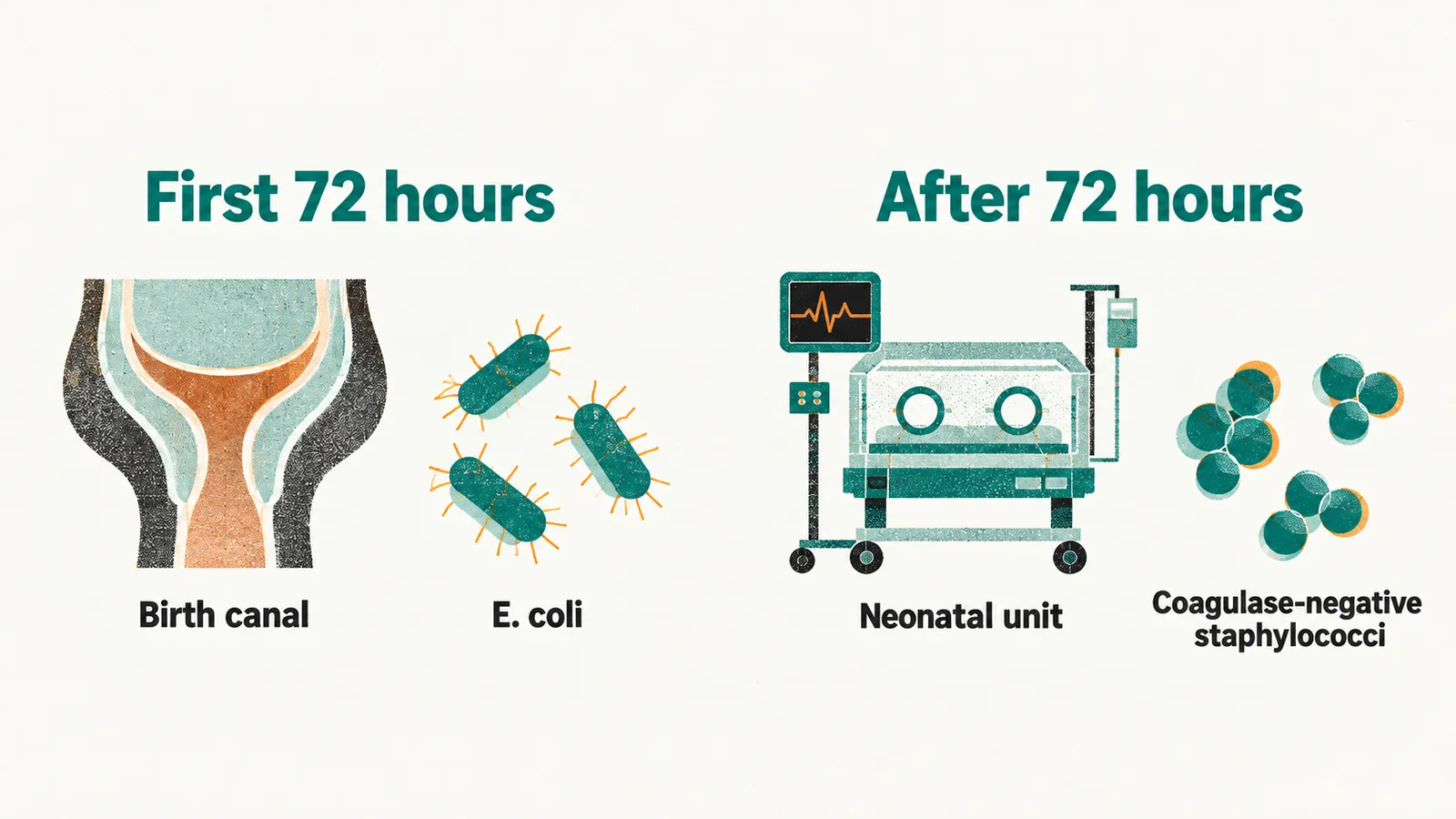
Neonatal infection means bacterial infection during the first 28 days of life, most importantly sepsis (infection of the bloodstream) and meningitis (infection of the cerebrospinal fluid). In a preterm infant the useful division is by timing rather than by organism: infection that appears in the first 72 hours is called early-onset, and infection appearing after that is called late-onset. The two groups differ in where the organisms come from, which organisms they are, and how they are treated.
Why prematurity is itself a risk factor
Prematurity raises the risk of infection in three ways: the infant’s own defenses are immature, the care the infant needs opens routes for bacteria, and the cause of the preterm birth may itself be infective.
- Antibody. Most maternal immunoglobulin G, the antibody class that crosses the placenta, does so in the third trimester, so an infant born before about 32 weeks receives little of the antibody repertoire that protects a term infant.
- White cells and complement. Neutrophil numbers and function, opsonization (the coating of bacteria that lets white cells engulf them) and complement activity are all reduced, so a bacterium that a mature immune system would contain can spread instead.
- Barriers. The skin is thin, easily macerated and repeatedly broken by tape, electrodes and needles; the gut mucosa is permeable; the airway is instrumented with a tube.
- Equipment. Central venous catheters, endotracheal tubes, parenteral nutrition and repeated blood sampling give organisms both a route into the circulation and a surface to colonize.
- The reason for the preterm birth. Preterm labor, preterm premature rupture of membranes (rupture before labor has begun) and chorioamnionitis (infection or inflammation of the fetal membranes) are themselves often infective or inflammatory, so infection is part of the differential from the first minutes of life rather than a complication that arrives later.
Early-onset infection comes from the birth canal
Early-onset sepsis is acquired before or during birth, typically from the maternal genital tract. A prospective network study of infants found an incidence of 13.5 per 1000 very preterm births (about 1.4%), with Escherichia coli accounting for 46.5% of the organisms and group B streptococcus for 18.8%. Group B streptococcus remains the more common cause in term infants, while E. coli predominates among preterm infants.
The clinical presentation is often respiratory: grunting, retractions, apnea, a rising oxygen requirement, or an infant who is difficult to stabilize after delivery. Risk factors in the history are ruptured membranes, maternal fever or chorioamnionitis, and known group B streptococcal colonization. Because early-onset infection can present as respiratory distress, it sits alongside respiratory distress syndrome in the differential of the first hours.
Late-onset infection comes from the unit
Late-onset sepsis is acquired after admission. Reported rates vary widely between units and are highest at the lowest gestational ages; in very-low-birth-weight infants, cohort studies commonly report roughly 15% to 25%. The organisms are different, because the source is different: coagulase-negative staphylococci are the most frequent isolates and are usually line-associated, followed by Gram-negative bacilli, Staphylococcus aureus and, in the most immature infants, Candida species.
Central venous catheters, mechanical ventilation, parenteral nutrition and long hospitalization all contribute, and the infections that follow are bloodstream infection, ventilator-associated pneumonia and sepsis arising from . Necrotizing enterocolitis and bloodstream infection frequently accompany each other, with a damaged gut wall allowing organisms into the circulation and infection worsening bowel perfusion, and the two can be difficult to separate at the bedside.
The presentation is non-specific
No single sign identifies neonatal sepsis in a preterm infant. The features that matter are the ones the infant cannot localize:
- Temperature instability, fever or, more often in a preterm infant, hypothermia.
- Increasing apnea, new respiratory distress or a rising oxygen requirement.
- Lethargy, hypotonia, poor perfusion, mottling, prolonged capillary refill or hypotension.
- Feeding intolerance, increasing gastric residuals, abdominal distension.
- Metabolic acidosis, unstable glucose, jaundice, or a sudden drop in platelet count.
Two consequences follow. Most infants who are investigated and treated for sepsis are not infected, and a preterm infant with proven sepsis often looks no different from several others in the same unit. The assessment therefore rests on repeated examination, on trends rather than single values, and on holding sepsis, necrotizing enterocolitis and metabolic decompensation in mind together, since they present with the same instability.
Making the diagnosis
Because the signs cannot confirm or exclude infection, the diagnosis rests on samples taken before treatment begins and on how the infant and the tests evolve afterwards.
Blood culture is the reference standard, and it should be taken before the first antibiotic dose. Volume matters: guidance asks for about 1 mL of blood from a small infant, because a smaller sample misses low-grade bacteremia. When a central line is in place, blood is taken from the line lumen as well as peripherally, because a line-associated infection may be detectable only there.
Alongside the culture, C-reactive protein, an inflammatory marker, is measured at the start and repeated at 18 to 24 hours in an infant who remains a concern, and the pair of values is more informative than either alone. It must not be used by itself to decide whether to start, continue or stop antibiotics, because it rises late and is raised by many non-infective causes.
Lumbar puncture is considered when there are signs suggesting meningitis, when infection is strongly suspected on clinical grounds, or when the blood culture is positive, because meningitis changes the choice and duration of treatment; in an unstable infant the decision is weighed against the risk of the procedure.
Treatment
Two principles govern the start of treatment. Antibiotics are given as soon as possible and always within 1 hour of the decision to treat, and the decision itself is not made for every infant with a risk factor, since most of them are not infected.
- Early-onset infection is treated empirically with intravenous benzylpenicillin plus gentamicin, the first choice in the United Kingdom guidance, with ampicillin plus gentamicin used in the same role in the United States; local resistance data override the default. The reference penicillin regimen is 25 mg/kg every 12 hours — the interval may be shortened to 8-hourly on clinical judgement — and gentamicin is started at 5 mg/kg. These are unit protocols, not doses to prescribe from a note; the exact dose and interval depend on gestational age, renal function and local policy.
- Late-onset infection in an infant already on a neonatal unit is treated with a narrow-spectrum combination that covers both Gram-positive and Gram-negative organisms, such as flucloxacillin plus gentamicin, chosen against local susceptibility data rather than by habit. If necrotizing enterocolitis is suspected, the regimen follows the unit’s gut-oriented protocol.
The course is then reviewed rather than simply continued. Current guidance reassesses the need for antibiotics at 36 hours for early-onset and 48 hours for late-onset infection. Treatment is stopped at that point if the blood culture is negative and the initial suspicion was not strong; it continues for 7 days if the culture is positive or if sepsis was strongly suspected despite a negative culture; and it is extended beyond 7 days when the infant has not recovered or the organism requires it. Meningitis is treated for longer than sepsis.
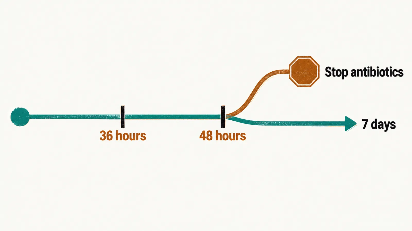
Stopping antibiotics is part of the treatment, not a failure of it. Every extra day of an unnecessary course selects resistant organisms, disturbs the developing intestinal microbiome, and is associated in cohort studies with necrotizing enterocolitis.
Preventing infection in the unit
Hand hygiene remains the most effective single measure. Beyond it, the aim is to reduce the number of ways into the bloodstream: central lines are inserted aseptically, reviewed every day and removed as soon as they are no longer needed, following the unit’s central line-associated bloodstream infection bundle; unnecessary procedures and unnecessary antibiotics are avoided; and feeding with human milk lowers the risk of necrotizing enterocolitis, which is one of the routes to late-onset sepsis.
Late-onset infection is a risk of the weeks in the unit rather than of the first days, and it is not the only one. Other injuries of prematurity also develop quietly over those weeks while the infant’s organs continue to mature outside the uterus, and some of them can be found only by looking for them on a schedule.