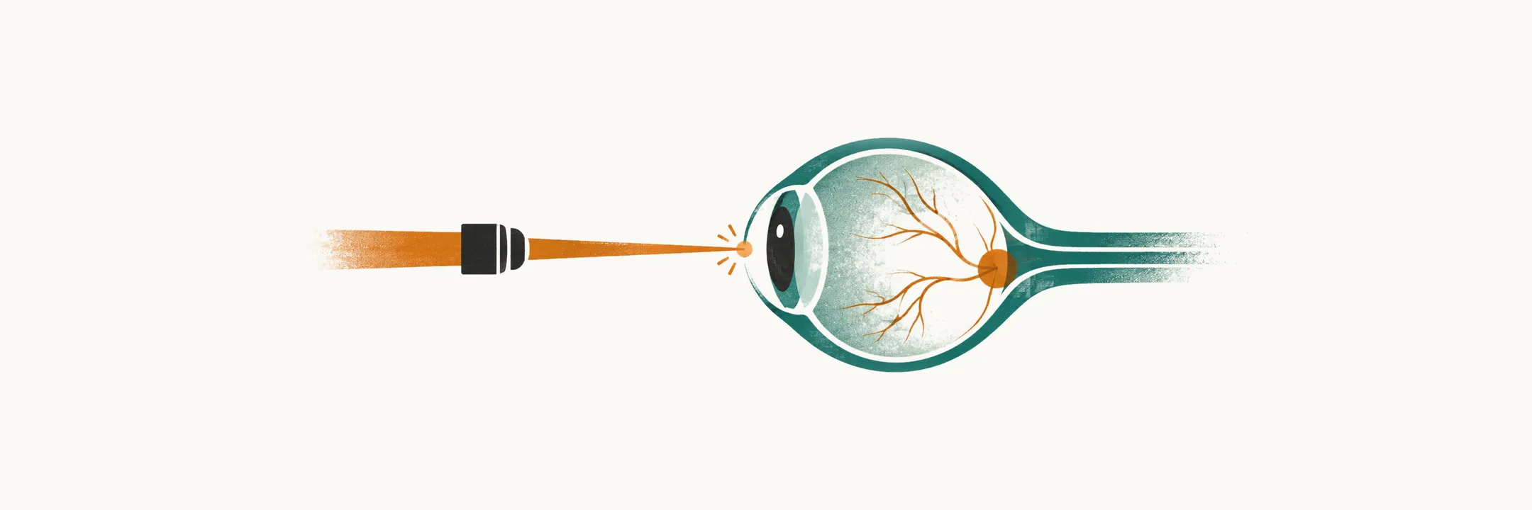
Retinopathy of prematurity (ROP) is abnormal development of the retinal blood vessels in an infant born before that vessel network has finished growing. It is not an injury to a mature retina; it is a disorder of vessels that were interrupted mid-growth, and it develops over weeks rather than hours. Untreated severe disease can detach the retina and cause blindness, which is why the disease is screened for by protocol instead of being looked for when vision is noticed to be poor.
The vessels that had not finished growing
The retina is vascularized from the optic disc outward. Vessels reach the nasal periphery by about 36 weeks of gestation but do not reach the temporal periphery until around term, so the temporal retina is the last part to become vascularized and the part most often left without a blood supply when an infant is born early. Where vessels have not grown, the inner retina is ischemic, and that ischemic tissue becomes the engine of the disease.
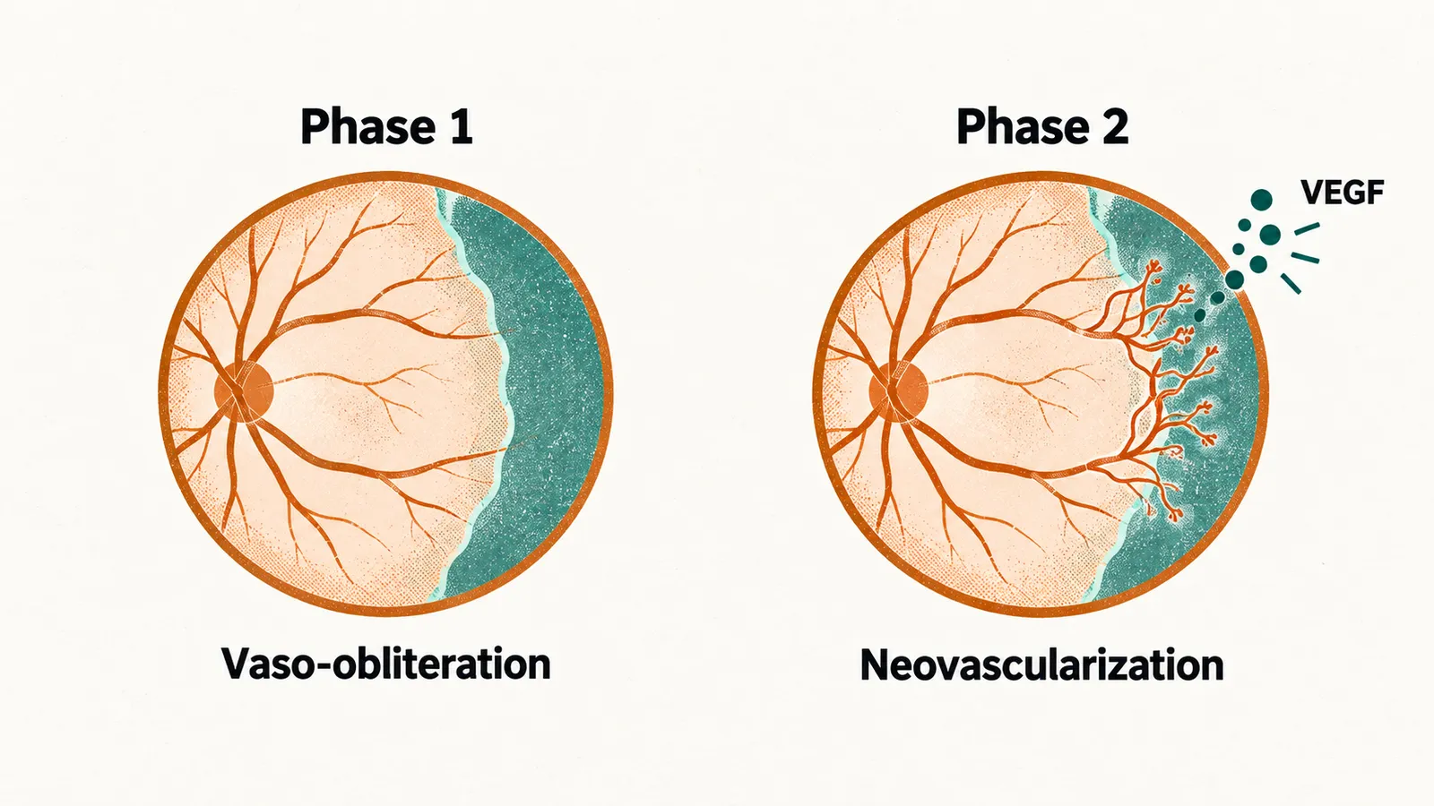
ROP runs through two phases that pull in opposite directions.
- Phase 1, vaso-obliteration. After birth the retina is exposed to an oxygen tension well above the fetal environment, even in room air, and it loses the placental growth factors it was receiving. Vessel growth stops or regresses, leaving a larger area of avascular retina than the birth alone would imply.
- Phase 2, neovascularization. The ischemic peripheral retina then releases vascular endothelial growth factor (VEGF), and new vessels form at the junction between vascularized and avascular retina. These vessels are abnormal: they leak, they bleed, and the fibrovascular tissue they build can contract and pull the retina off the underlying wall.
Oxygen therefore sits on both sides of the disease. Too much arrests vessel growth and creates the ischemia; too little drives the second, proliferative phase. The same tension shapes the saturation target used in the delivery room, where oxygen is titrated to a protocol-defined range in The Golden Hour and NICU Care of the Preterm Infant, because both extremes cause harm.
Oxygen targets: a trade-off with no comfortable answer
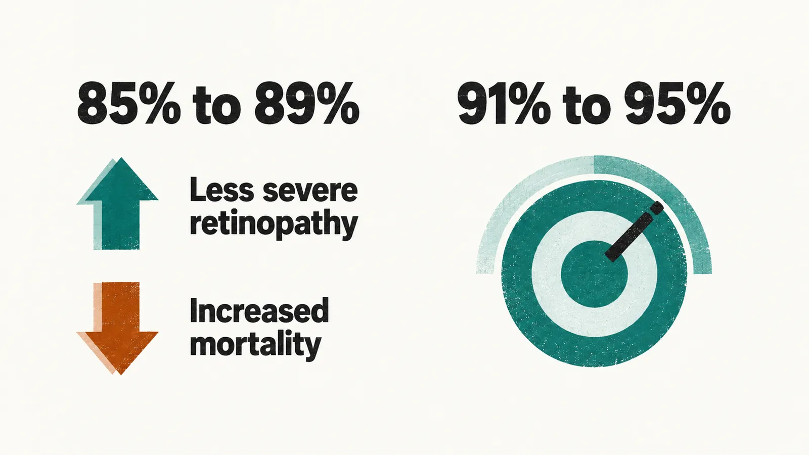
Because oxygen is both necessary and injurious to the developing retina, several large trials compared a lower saturation target (85% to 89%) with a higher one (91% to 95%) in infants. The lower target reduced severe retinopathy but increased mortality, and the pooled analysis of those trials confirmed that the two cannot be separated. No single target has therefore been adopted as safe: units choose a range, commonly about 90% to 95%, and rely on alarm limits, nursing vigilance and avoidance of both sustained hyperoxemia and hypoxemia. Oxygen saturation targeting is an example of a setting where the population-level optimum is a compromise rather than a value that is safe for every infant.
How the disease is described
Because the disease begins at the border between vascularized and avascular retina, an examination describes where that border lies, what it looks like and how active the retinal vessels are. Findings are recorded by , and vascular activity, using the international classification.
- Zone records how posterior the disease is: zone I surrounds the optic disc and macula, zone II extends to the nasal periphery, and zone III is the temporal crescent that vascularizes last. Posterior disease is the more dangerous.
- Stage records the junction appearance: stage 1, a flat demarcation line; stage 2, a raised ridge; stage 3, extraretinal fibrovascular proliferation into the vitreous; stage 4, partial retinal detachment; stage 5, total retinal detachment.
- describes increased venous tortuosity and arterial dilatation in the posterior pole, the central back of the retina around the optic disc and macula. It is a marker of severe vascular activity rather than a stage, and its presence changes the urgency.
- Aggressive posterior ROP, previously called rush disease, is a rapidly progressive posterior form, most often seen in the least mature infants, in which the usual staging sequence is compressed.
Screening finds it before it can be seen from outside
An infant cannot report failing vision, and ROP is invisible from the outside until a retinal detachment is advanced, so screening is defined by risk rather than by symptoms. The screening population is infants with a birth weight of 1500 g or less or a gestational age of 30 weeks or less, plus selected heavier or more mature infants in whom the neonatal course was unusually unstable, at the judgement of the clinical team. Pooled population-based estimates put any retinopathy at about 30% of very-low-birth-weight infants and severe disease at about 9%, so the programme examines many infants in order to find the minority who need treatment.
The first examination is timed by maturity. For infants born at 22 to 27 weeks it is due at about 31 weeks ; for those born at 28 to 30 weeks it follows at 32 to 34 weeks postmenstrual age, or at 4 weeks of chronological age, whichever is later. Examinations then continue at intervals set by the findings, and screening ends only when the temporal retina is fully vascularized or the disease has clearly regressed. The examination is done by an ophthalmologist with indirect ophthalmoscopy, with the pupils dilated in advance and the infant monitored during and after the procedure.
When treatment is needed
Most ROP regresses on its own. Treatment is directed at the group with : any stage in zone I with plus disease, stage 3 in zone I without plus disease, and stage 2 or 3 in zone II with plus disease. This threshold is treated promptly, generally within 72 hours of diagnosis, because disease at this point can progress to retinal detachment within days.
Two treatments are in use.
- Laser photocoagulation of the avascular peripheral retina removes the ischemic tissue that is driving VEGF production, and has been the standard treatment since trials showed that treating type 1 disease early preserves vision better than observing until threshold disease.
- Intravitreal anti-VEGF injection blocks the growth factor directly. It produces faster regression and is often used for posterior disease, where laser treatment is technically difficult. Its drawback is that the effect is temporary: reactivation can occur later, after the usual screening period, so infants treated with anti-VEGF need prolonged ophthalmological surveillance.
Stage 4 and stage 5 disease, with retinal detachment, requires vitreoretinal surgery, and the visual outcome is much less predictable than after timely treatment of type 1 disease.
After the acute disease
Regression is the usual outcome of treated type 1 disease, but it is not the end of the story. Children who have had any ROP, including disease that resolved without treatment, carry a higher risk of myopia, strabismus and refractive error, so ophthalmological follow-up continues well beyond the neonatal period. Treatment of severe disease markedly reduces the blindness it would otherwise cause; the disease remains a leading cause of childhood blindness where screening or treatment is unavailable, which is the practical argument for protocol-driven examination of every infant who meets the risk criteria.