
A patent ductus arteriosus (PDA) is persistence after birth of the fetal vessel that connects the pulmonary artery to the descending aorta. Functional closure normally begins within hours of birth and anatomical closure follows over days to weeks. In a preterm infant the ductus often remains open because the signals that close it — lung aeration, rising oxygen tension, falling prostaglandin exposure and reversal of the fetal pressure gradient — are weak or delayed.
What the ductus does before birth
Fetal pulmonary vascular resistance is high because the lungs are fluid-filled and do not perform gas exchange. Blood entering the pulmonary artery therefore bypasses the lungs through the ductus and flows right to left into the aorta, and oxygenation happens in the placenta.
At birth two changes occur together. Clamping the cord removes the low-resistance placental circulation and raises systemic vascular resistance; aeration of the lung lowers pulmonary vascular resistance. Flow across the ductus first reverses to left to right, and the vessel then constricts as oxygen tension rises and placental prostaglandin input falls.
Why the ductus stays open in prematurity
Three mechanisms reinforce each other:
- Low oxygen tension. Respiratory distress syndrome limits oxygenation, and hypoxia is a potent ductal vasodilator, so the constriction signal is weak.
- Persistent prostaglandin activity. The ductus is held open by prostaglandin E2 and prostacyclin; the immature lung metabolizes and clears these mediators more slowly, so vasodilator tone persists.
- Delayed fall in pulmonary vascular resistance. Poor lung inflation keeps pulmonary pressures high, delaying the pressure reversal that normally accompanies the change in flow direction.
Lung disease therefore promotes ductal patency, and an open ductus then increases pulmonary blood flow and worsens lung edema, making respiratory support more difficult. Spontaneous closure is common but not the rule at the earliest gestational ages: in infants managed without intended closure, roughly one third close in the first one to two weeks, with further closures over subsequent weeks.
Flow direction determines the clinical problem
Once the ductus stays open, blood crosses it from whichever side has the higher pressure, and each direction of this shunt produces a different problem.
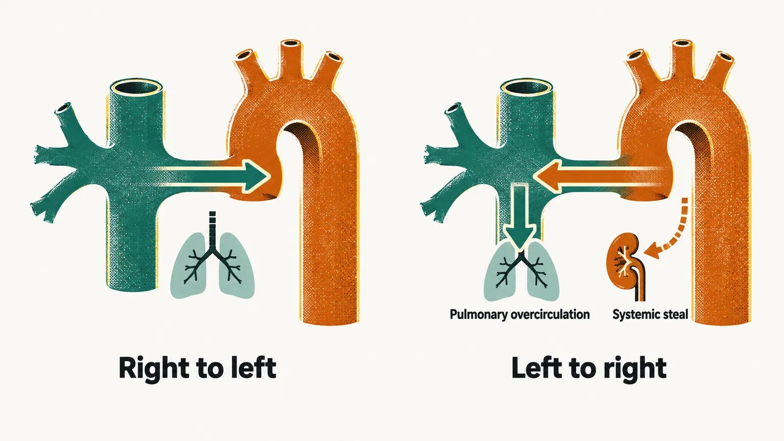
Right-to-left shunting persists when pulmonary pressure remains above systemic pressure. Blood bypasses the lungs and systemic saturation falls distal to the ductus. Oxygen saturation measured on the right arm (preductal, supplied by blood that has not passed the ductus) is compared with a lower limb or left arm (postductal), and a difference suggests persistent fetal-type shunting or pulmonary hypertension.
Left-to-right shunting develops when systemic pressure exceeds pulmonary pressure while the ductus stays open. Blood runs from the aorta into the pulmonary artery, producing pulmonary overcirculation, pulmonary edema and increased pulmonary venous return to the left atrium, which can enlarge and eventually fail.
Left-to-right flow can also systemic blood during diastole, because blood escapes into the lower-pressure pulmonary circulation when the systemic circulation should be perfusing organs. Doppler studies, which use ultrasound to measure blood flow, may show absent or reversed diastolic flow in systemic vessels. This is the hemodynamic link between a hemodynamically significant PDA and renal dysfunction, reduced gut perfusion and cerebral vulnerability, including the ischemic contribution to necrotizing enterocolitis.
Diagnosis is hemodynamic, not merely anatomical
Clinical signs — a systolic murmur, a bounding pulse, a wide pulse pressure, hyperactive precordium — appear late and are unreliable in small infants. Echocardiography is the standard tool: it confirms the ductus, identifies the direction of flow, measures the ductal diameter, and estimates whether the shunt is hemodynamically significant through effects such as left atrial enlargement and altered diastolic flow in systemic vessels.
The question that matters at the bedside is not simply whether the ductus is open, but whether it is worsening pulmonary disease or reducing systemic organ perfusion enough to justify closing it. A small ductus in a stable infant is often observed.
Closing the ductus: benefit against organ toxicity
Supportive measures come first: optimizing ventilation and lung recruitment lowers pulmonary vascular resistance and strengthens the oxygen signal for constriction, and fluid and diuretic management limits pulmonary congestion. Oxygen is deliberately not pushed high, because hyperoxia contributes to .
Prostaglandin synthesis inhibition is the pharmacological route, using a cyclooxygenase inhibitor, a drug that blocks the enzyme that makes prostaglandins.
- Ibuprofen is widely used, in the regimen of 10 mg/kg followed by 5 mg/kg at 24 hours and again at 48 hours. This is a standard neonatal protocol, not a prescription to dose outside a unit’s own policy.
- Indomethacin has similar efficacy but a less favorable profile in pooled trials, with more transient renal impairment and oliguria, so ibuprofen is often preferred.
- Paracetamol (acetaminophen) works through a different mechanism and, in pooled evidence, achieves closure rates similar to ibuprofen with fewer renal and intestinal adverse effects, although the intravenous route has been less effective than the enteral route in some trials and evidence quality remains limited. It is often considered when bleeding risk, thrombocytopenia, renal risk or bowel disease makes cyclooxygenase inhibitors unsuitable.
The trade-off is real and must be stated plainly: a drug that closes the ductus reduces pulmonary overload but can worsen renal and bowel perfusion, especially at the lowest gestational ages. is the sharpest conflict, because indomethacin and ibuprofen may worsen bowel ischemia or perforation. When active necrotizing enterocolitis is present or strongly suspected, closure is deferred and the open ductus is managed supportively, with fluids and diuretics, until the bowel recovers.
Surgical ligation is reserved for infants in whom medical closure has failed or is contraindicated, or who remain ventilator-dependent because of the shunt. It closes the vessel immediately, but that sudden change forces the immature circulation to adapt in seconds rather than over days, and postoperative hemodynamic instability is the main concern.
Whichever route is chosen, the ductus is not only a heart problem. Its steal already links it to renal dysfunction, reduced gut perfusion and cerebral vulnerability, and the brain, whose regulation of its own blood flow is still immature, is poorly protected against swings in blood pressure.