Survival does not end the disease of prematurity. A preterm infant, born before 37 completed weeks of gestation, may move from acute respiratory and circulatory failure into weeks or months of lung support, feeding dependence, infection risk, sensory injury and neurodevelopmental vulnerability. Follow-up has two jobs: track the organ injuries accumulated during intensive care, and interpret later growth and development using , which counts age from the expected due date instead of from birth, rather than chronological age alone.
Complications cluster at different postnatal times
The timeline of mortality and morbidity follows the order in which immature organs meet the demands of extrauterine life.
- The first hours. Profound whole-body immaturity may be incompatible with life outside the uterus, and the transition itself can fail.
- The first two weeks. Respiratory distress syndrome, the lung failure caused by surfactant deficiency, becomes a major cause of death, together with the circulatory transition and early brain injury.
- The following weeks. (inflammatory necrosis of the immature bowel), hospital-acquired infection and the consequences of prolonged intensive care dominate.
- Beyond the acute admission. Chronic lung disease can remain a life-threatening respiratory condition, and neurodevelopmental, visual and growth problems become apparent over months and years.
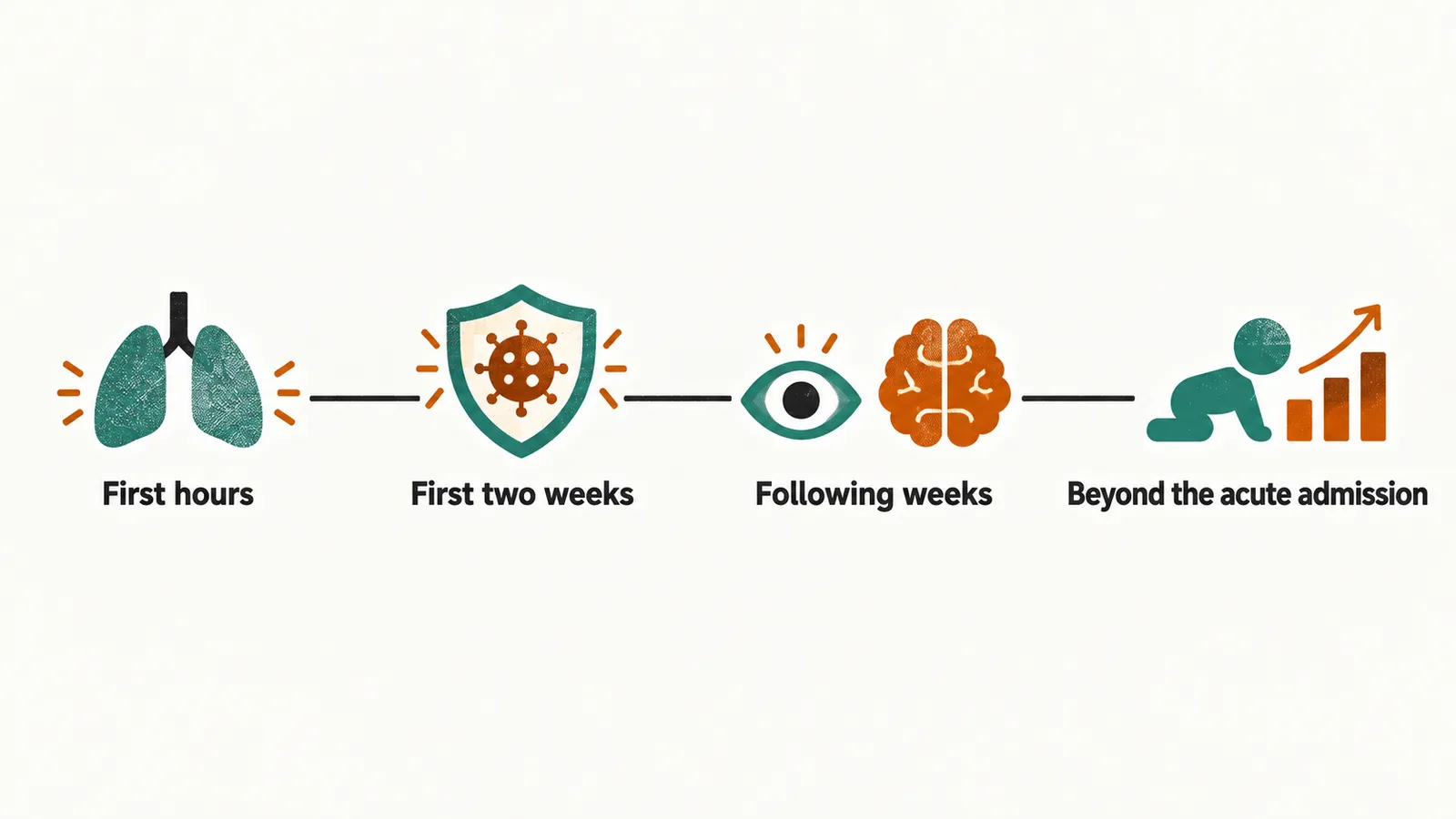
Because each interval has its own substrate, the question “what is this infant at risk of now?” has a different answer in week one than in month three.
Bronchopulmonary dysplasia
(BPD), also called chronic lung disease of prematurity, is the best-characterized chronic complication after birth. It develops in a lung that is still trying to grow while being exposed to oxygen, inflammation, ventilator stress and fluid overload. Definitions differ, so incidence depends on which one is used; among extremely preterm infants surviving to 36 weeks (gestational age at birth plus the weeks since), about half meet an oxygen-use definition.
Affected infants may stay on respiratory support and saturation monitoring for weeks or months, feed late because breathing consumes their energy budget, and in severe cases go home with oxygen or, rarely, a tracheostomy and long-term ventilatory support. Imaging ranges from fine granular opacity in moderate disease to coarse interstitial change, hyperinflation, fibrosis or persistent ventilation in severe disease.
What the child may carry forward
The lung is the most visible legacy, but it is not the only one. Most of the morbidity that matters later is established during the neonatal weeks and then expresses itself over years, which is why one child can be followed for lung, gut, brain and eye consequences at the same time. Two of these decide much of the long-term picture.
Brain injury is the substrate of most later cerebral palsy among preterm survivors, and its frequency falls as gestational age rises. It includes germinal matrix hemorrhage, bleeding from a fragile vascular region beside the ventricles; , usually a venous infarction of the brain tissue beside the ventricle; and , small cysts left by injury to the periventricular white matter. The most immature survivors carry the highest risk of spastic motor impairment, and they also carry a higher risk of cognitive, language and behavioral difficulty, which does not show itself in the same obvious way.
, abnormal growth of retinal vessels that had not finished developing at birth, may leave myopia, strabismus or refractive error even when the acute disease resolved, so the eyes still need review after discharge. It remains a leading cause of childhood blindness where screening and treatment are not available, and treatment of severe disease markedly reduces that outcome.
Growth and feeding
Feeding autonomy is often the last milestone of the neonatal admission. Respiratory support, immature suck–swallow coordination and the increased work of breathing in BPD all interfere with oral feeding, and bowel resection after necrotizing enterocolitis can reduce absorptive surface. Growth therefore cannot be interpreted without the lung, gut and neurological history.
Growth and development are interpreted using corrected age (Corrected Age and Clinical Risk in the Preterm Infant): a child born 12 weeks early will reach some milestones later by chronological age even when development is appropriate for corrected age, and age correction for growth in extremely and children may be needed out to 36 months.
Follow-up and the family
Follow-up programmes monitor the domains that can fail silently: respiratory symptoms and lung function where relevant, growth, feeding and nutrition, hearing, vision, motor development, and language and cognition. Exactly which tests are done, and at what intervals, follows local programme protocol, because the published schedules differ and thresholds change with evolving evidence.
The family is part of that monitoring. The prolonged admission changes family life: parents may be separated from work, home and other children for months, and they were often prepared for a healthy term newborn rather than an infant on respiratory support. Family-centered care keeps them involved during the admission; after discharge they are the people who observe breathing, feeding, growth and development every day, and their reports are clinical information.