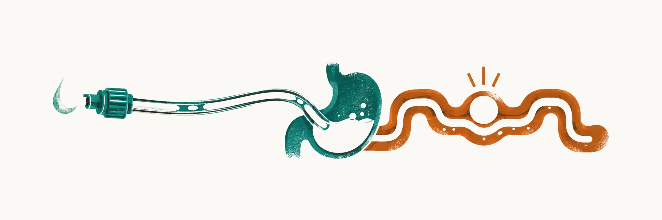
Feeding a preterm infant is a developmental problem before it is a calorie problem. The infant needs substrates for rapid growth, but the coordination required to feed and the tolerance of the immature intestine are both incomplete. The plan therefore combines human milk, tube feeding, cautious progression of enteral volume and intravenous nutrition, while watching for necrotizing enterocolitis (NEC) — inflammatory necrosis of the immature bowel.
Why the infant cannot simply breastfeed
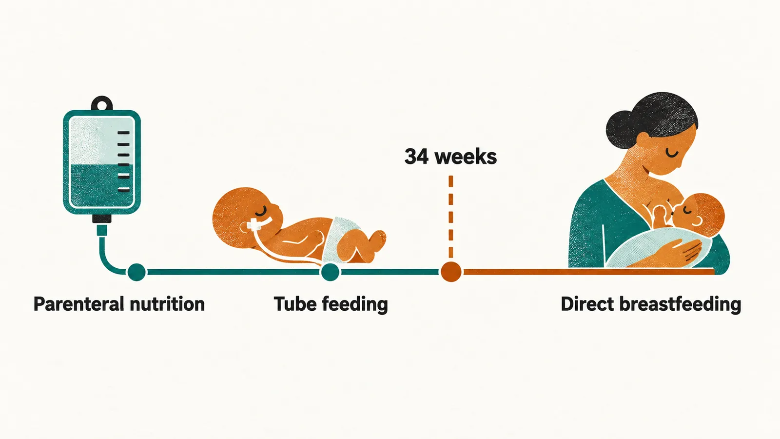
Coordinated sucking, swallowing and breathing is not reliably mature until about 34 weeks . Younger infants may suck non-nutritively but cannot transfer a full feed safely, because the sequence of suck, swallow and breathe can become discoordinated and lead to aspiration or desaturation. This is why a usually cannot breastfeed directly even when human milk is strongly preferred.
The mother expresses milk, which is given through a nasogastric or orogastric tube; when maternal milk is not available, donor human milk is the preferred substitute. The route is adapted to maturity while the nutritional substrate stays as close as possible to human milk. Human milk feeding is associated with lower NEC risk than formula in preterm infants, which is one reason it is preferred when it is available.
The intestine is immature in parallel: motility, enzyme activity, mucosal barrier function and the ability to tolerate a full enteral (into the gut) volume all develop after birth. Practice therefore combines parenteral nutrition, given intravenously, with tiny early enteral feeds, often called minimal (trophic) enteral feeding, and then increases enteral volume slowly. The small feeds expose the gut to milk and support mucosal adaptation, while intravenous nutrition supplies what the gut cannot yet absorb safely.
Vascular access supports the transition
The intravenous part of this plan depends on reliable access to a vein. Immediately after birth the umbilical vein offers rapid central access for fluids and parenteral nutrition. An umbilical venous catheter is usually kept for about five to seven days, because the risk of bloodstream infection rises with dwell time; many units follow a recommendation to remove it at or before seven days if it is still needed, and insert a percutaneously inserted central catheter, a thin line passed through a peripheral vein into a central vein, for prolonged support. The indication and duration of every line are reviewed daily, because each one adds infection and vascular risk.
What necrotizing enterocolitis is
NEC is an acute inflammatory disease in which the bowel wall becomes injured and necrotic, most often in the terminal ileum and proximal colon. Its cause is multifactorial, and feeding is a contributor rather than the cause:
- The immature mucosal barrier and immune response react abnormally to enteral substrate.
- Antibiotic exposure and other factors alter the developing intestinal microbiome, the community of microorganisms that colonizes the gut.
- Infection and dysbiosis, a disturbed balance of that community, amplify inflammation.
- Reduced bowel perfusion, including , in which blood is diverted away from the organs through a hemodynamically significant patent ductus arteriosus, makes the bowel wall more vulnerable to ischemic injury.
NEC affects roughly 7% to 10% of very-low-birth-weight infants, and reported mortality is in the region of 20% to 30% overall, with pooled figures around 35% for infants needing surgery and about 50% in infants with surgical disease. Those figures vary by population and era, but they explain why prevention and early recognition dominate feeding decisions in the neonatal unit.
Severe intestinal inflammation is not confined to the abdomen. Shock, acidosis and circulating inflammatory mediators are associated with white-matter injury, which is one reason NEC matters well beyond the bowel.
Recognition: local abdominal clues against systemic deterioration
Abdominal distension, increasing gastric residuals (milk still in the stomach when the next feed is due), bilious (bile-stained) drainage from the feeding tube, blood in the stool, abdominal tenderness and abdominal wall discoloration all point to bowel disease. The difficulty is that preterm infants often show non-specific systemic signs first: temperature instability, more frequent apnea, lethargy, poor perfusion, hypotension, metabolic acidosis, respiratory failure or shock.
That overlap creates the differential a clinician has to hold in mind: sepsis, NEC and metabolic disease can all present as an infant who has suddenly become less stable, and they may coexist. The abdomen and imaging help separate them, but cultures, blood tests and repeated examination remain necessary.
Imaging: pneumatosis is the hallmark
Abdominal radiography may show , gas within the bowel wall, which is the radiographic hallmark of NEC. Gas may also appear in the portal venous system. Free intraperitoneal air indicates perforation and converts the illness into a surgical emergency.
Staging systems such as the separate suspected disease, definite disease with characteristic intestinal signs, and advanced disease with systemic deterioration, shock or perforation. The practical learning boundary is that distinction rather than every numbered substage: it corresponds to escalation of treatment and to the decision about surgery.
Immediate treatment follows the mechanism
Treatment addresses each part of the disease at once: the injured bowel, the possibility of infection and the systemic illness.
- Stop enteral feeding, so the injured bowel is no longer challenged metabolically.
- Switch nutrition to total parenteral support while enteral feeding is held.
- Start broad-spectrum antibiotics while cultures are pending, because infection may be the cause, a consequence, or clinically indistinguishable from NEC.
- Support circulation and ventilation, treating shock, acidosis and respiratory failure.
- Reassess repeatedly, because deterioration can be rapid and progression to perforation is the event that must not be missed.
Perforation requires drainage or resection of necrotic bowel. Extensive resection leaves infants at risk of later strictures and short-bowel syndrome, which is why the aim is to stop progression before tissue is lost.
Treatment can also collide with care of the ductus. Indomethacin and ibuprofen close the ductus by inhibiting prostaglandin synthesis, but they also reduce bowel and renal perfusion. In the presence of active NEC they may worsen ischemia or perforation, so closure is deferred and the patent ductus is managed supportively instead. The team may have to accept an open ductus temporarily rather than treat the heart at the bowel’s expense.
Infection stays close to the diagnosis throughout. A damaged bowel wall can let organisms into the circulation, and bloodstream infection can be clinically indistinguishable from NEC, which is why cultures are taken and antibiotics started before the two can be told apart.